. 2018 May 2;9(1):124.
doi: 10.1186/s13287-018-0863-8.

Human platelet lysate in mesenchymal stromal cell expansion according to a GMP grade protocol: a cell factory experience

Affiliations

Human platelet lysate in mesenchymal stromal cell expansion according to a GMP grade protocol: a cell factory experience

Valentina Becherucci et al. Stem Cell Res Ther. .

Abstract

Background: The use of platelet lysate (PL) for the ex-vivo expansion of mesenchymal stromal/stem cells (MSCs) was initially proposed by Doucet et al. in 2005, as an alternative to animal serum. Moreover, regulatory authorities discourage the use of fetal bovine serum (FBS) or other animal derivatives, to avoid risk of zoonoses and xenogeneic immune reactions. Even if many studies investigated PL composition, there still are some open issues related to its use in ex-vivo MSC expansion, especially according to good manufacturing practice (GMP) grade protocols.

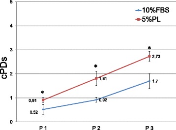
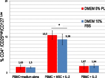
Methods: As an authorized cell factory, we report our experience using standardized PL produced by Azienda Ospedaliero Universitaria Meyer Transfusion Service for MSC expansion according to a GMP grade clinical protocol. As suggested by other authors, we performed an in-vitro test on MSCs versus MSCs cultured with FBS that still represents the best way to test PL batches. We compared 12 MSC batches cultured with DMEM 5% PL with similar batches cultured with DMEM 10% FBS, focusing on the MSC proliferation rate, MSC surface marker expression, MSC immunomodulatory and differentiation potential, and finally MSC relative telomere length.

Results: Results confirmed the literature data as PL increases cell proliferation without affecting the MSC immunophenotype, immunomodulatory potential, differentiation potential and relative telomere length.

Conclusions: PL can be considered a safe alternative to FBS for ex-vivo expansion of MSC according to a GMP grade protocol. Our experience confirms the literature data: a large number of MSCs for clinical applications can be obtained by expansion with PL, without affecting the MSC main features. Our experience underlines the benefits of a close collaboration between the PL producers (transfusion service) and the end users (cell factory) in a synergy of skills and experiences that can lead to standardized PL production.

Keywords: Advanced therapy medicinal products; Cell factory; Fetal bovine serum; Good manufacturing practice; Mesenchymal stromal/stem cells; Platelet lysate.


Conflict of interest statement

Ethics approval and consent to participate

Not applicable.

Competing interests

The authors have no commercial, proprietary or financial interest in the products or companies described in this article.

Publisher’s Note

Springer Nature remains neutral with regard to jurisdictional claims in published maps and institutional affiliations.

Figures

Fig. 1
Fig. 1
MSC cumulative population doublings calculated from P1 to P3 in culture conditions DMEM 10% FBS versus DMEM 5% PL. n = 12. Data presented as mean ± SD. *p < 0.05. cPD cumulative population doubling, FBS fetal bovine serum, PL platelet lysate
Fig. 2
Fig. 2
Morphological changes induced by PL (representative case). a When observed by inverted microscope (10×), MCSs cultivated in presence of PL appear smaller, more refractive and less attached to plastic support than MCSs cultured with FBS. b Morphological changes confirmed by flow cytometry analysis of FSC and SSC, as cells cultured in PL showed reduction of FSC and SSC rate representative of less complexity and smaller dimensions. DMEM Dulbecco’s modified Eagle medium, FBS fetal bovine serum, FSC forward scatter, MSC mesenchymal stromal/stem cell, PL platelet lysate, SSC side scatter
Fig. 3
Fig. 3
MSC differentiation potential assays. MSC differentiation potential assay after 15 or 21 days of specific induction in both culture conditions. a, b Alcian Blue staining shows hyaluronic acid for chondrocytes, c, d Oil Red O shows intracytoplasmatic vacuoles in adipocytes and e, f Alizarin Red S staining shows presence of calcium matrix in osteoblasts, respectively, in PL-MSCs and FBS-MSCs. n = 12. FBS fetal bovine serum, LP platelet lysate
Fig. 4
Fig. 4
Induction of T-regulatory cell population by MSCs cultured in PL or FBS-containing medium. Data presented as mean ± SD with n = 10. Treg cell induction by MSCs evaluated as percentage of CD25High/CD4+/CD127Low/− PBMCs after 7 days of coculture. No significant differences found between MSCs cultured in PL or FBS-containing medium. In both culture conditions at day 7, percentage of CD4+/CD25high/CD127Low/− T cells was significantly higher (*p < 0.05) in cocultures with MSCs compared to PBMCs alone. DMEM Dulbecco’s modified Eagle medium, FBS fetal bovine serum, IL interleukin, MSC mesenchymal stromal/stem cell, PBMC peripheral blood mononuclear cell, PL platelet lysate
Fig. 5
Fig. 5
Inhibition of MLR by MSCs cultured in PL or FBS-containing medium. Data presented as mean ± SD with n = 10. Inhibition of PMBC responder (R) proliferation in presence of both allogeneic (PBMC stimulator (S)) and polyclonal (aCD3/28) stimuli evaluated as percentage of proliferation (% CFDA-SE-positive cells) in presence/absence of MSCs on 7-AAD CD45/CD3+ cells. No significant differences found between MSCs cultured in PL or FBS-containing medium. In both culture conditions, MSCs significantly reduce PBMC proliferation compared to PBMCs alone (*p < 0.05) in a dose-dependent manner. FBS fetal bovine serum, MSC mesenchymal stromal/stem cell, PL platelet lysate
Fig. 6
Fig. 6
MSC relative telomere length at passage 4. Data presented as mean ± SD with n = 12. RTL value calculated as ratio between telomere signal of each sample and control cells (K562 line) with correction for DNA index of G0–G1 cells. No significant differences observed (p > 0.05). FBS fetal bovine serum, NS not significant, PL platelet lysate

References

    1. Bobis S, Jarocha D, Majka M. Mesenchymal stem cells: characteristics and clinical applications. Folia Histochem Cytobiol. 2006;44(4):215–230. - PubMed
    1. Rosenbaum AJ, Grande DA, Dines JS. The use of mesenchymal stem cells in tissue engineering: a global assessment. Organ. 2008;4(1):23–27. - PMC - PubMed
    1. Ullah I, Subbarao RB, Rho GJ. Human mesenchymal stem cells—current trends and future prospective. Biosci Rep. 2015;35(2) 10.1042/BSR20150025. - PMC - PubMed
    1. Miao C, Lei M, Hu W, Han S, Wang Q. A brief review: the therapeutic potential of bone marrow mesenchymal stem cells in myocardial infarction. Stem Cell Res Ther. 2017;8(1):242. doi: 10.1186/s13287-017-0697-9. - DOI - PMC - PubMed
    1. Cui D, Li H, Xu X, Ye L, Zhou X, Zheng L, Zhou Y. Mesenchymal stem cells for cartilage regeneration of TMJ osteoarthritis. Stem Cells Int. 2017;2017:5979741. - PMC - PubMed

LinkOut - more resources