. 2018 Jun 29;293(26):9958-9969.
doi: 10.1074/jbc.RA118.003176. Epub 2018 May 2.

Crystal structures of the human 4-1BB receptor bound to its ligand 4-1BBL reveal covalent receptor dimerization as a potential signaling amplifier

Affiliations

Crystal structures of the human 4-1BB receptor bound to its ligand 4-1BBL reveal covalent receptor dimerization as a potential signaling amplifier

Aruna Bitra et al. J Biol Chem. .

Abstract

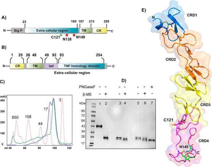
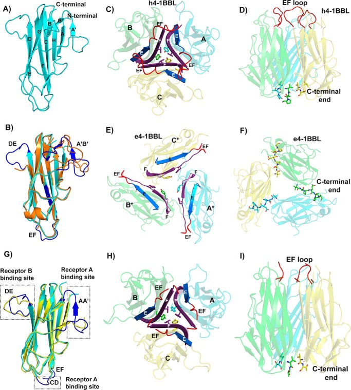
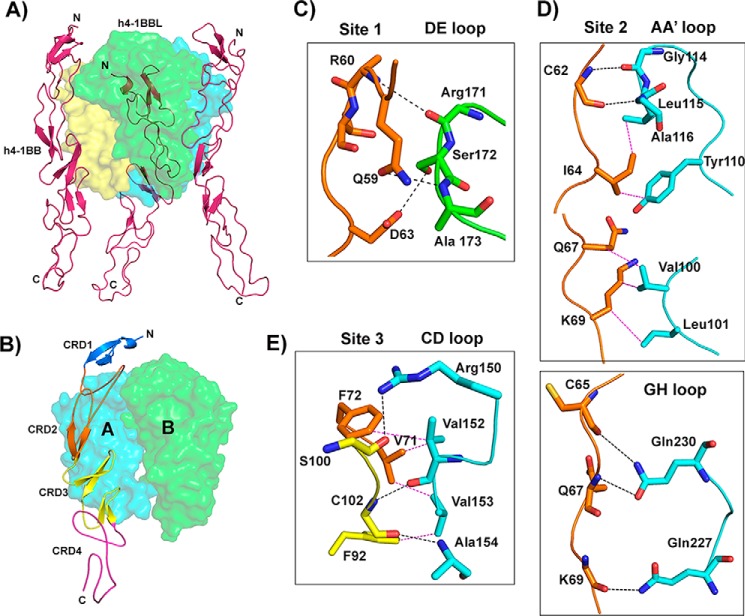
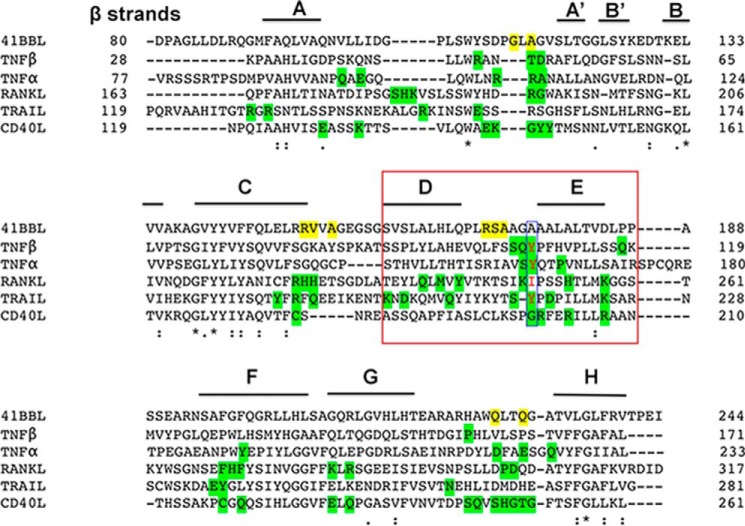
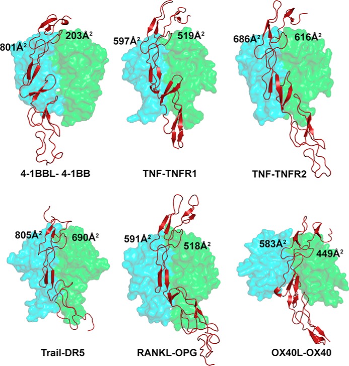
Human (h)4-1BB (TNFRSF9 or CD137) is an inducible tumor necrosis factor receptor (TNFR) superfamily member that interacts with its cognate ligand h4-1BBL to promote T lymphocyte activation and proliferation. h4-1BB is currently being targeted with agonists in cancer immunotherapy. Here, we determined the crystal structures of unbound h4-1BBL and both WT h4-1BB and a dimerization-deficient h4-1BB mutant (C121S) in complex with h4-1BBL at resolutions between 2.7 and 3.2 Å. We observed that the structural arrangement of 4-1BBL, both unbound and in the complex, represents the canonical bell shape as seen in other similar TNF proteins and differs from the previously reported three-bladed propeller structure of 4-1BBL. We also found that the binding site for the receptor is at the crevice formed between two protomers of h4-1BBL, but that h4-1BB interacts predominantly with only one ligand protomer. Moreover, h4-1BBL lacked the conserved tyrosine residue in the DE loop that forms canonical interactions between other TNFR family molecules and their ligands, suggesting h4-1BBL engages h4-1BB through a distinct mechanism. Of note, we discovered that h4-1BB forms a disulfide-linked dimer because of the presence of an additional cysteine residue found in its cysteine-rich domain 4 (CRD4). As a result, h4-1BB dimerization, in addition to trimerization via h4-1BBL binding, could result in cross-linking of individual ligand-receptor complexes to form a 2D network that stimulates strong h4-1BB signaling. This work provides critical insights into the structural and functional properties of both h4-1BB and h4-1BBL and reveals that covalent receptor dimerization amplifies h4-1BB signaling.

Keywords: X-ray crystallography; cell surface receptor; protein structure; protein-protein interaction; recombinant protein expression; tumor necrosis factor (TNF).


Conflict of interest statement

The authors declare that they have no conflicts of interest with the contents of this article

Figures

Figure 1.
Figure 1.
Protein constructs, expression, and h4-1BB structure. A and B, domain architecture of h4-1BB (A) and h4-1BBL (B). Sig p, signal peptide; TM, transmembrane region; CR, cytoplasmic region. The unpaired cysteine 121 and two potential N-linked glycosylation sites of h4-1BB are indicated. C, size exclusion profile of purified wildtype (WT) (blue line) and C121S mutant (green line) of h4-1BB with reference to molecular mass marker proteins (red line) in kDa. D, SDS-PAGE analysis of purified WT and C121S mutant of h4-1BB under nonreducing (−β-ME (lanes 1, 3, and 5) and reducing conditions (+β-ME (lanes 2, 4, and 6). Lanes 1 and 2 contain dimeric WT 4-1BB (blue high Mr peak from C) and lanes 3 and 4 contain monomeric WT 4-1BB (blue low Mr peak from C). Lanes 5 and 6 contain the C121S mutant 4-1BB (green low Mr peak from C). Lane 7 represents deglycosylated (PNGase (peptide N-glycosidase F) treated) WT 4-1BB. E, crystal structure of h4-1BB (in the 4-1BB–4-1BBL complex) colored by the four cysteine-rich domains as cartoon overlaid onto transparent molecular surface representation. Unpaired Cys121 and disulfide bridges are shown as sticks in respective colors of CRDs. The N-linked glycosylation site and the N-glycans are represented as sticks with carbon atoms in green, oxygen in red, and nitrogen atoms in blue.
Figure 2.
Figure 2.
Structure of h4-1BB ligand in the h4-1BB–4-1BBL complex. A, cartoon representation of the THD region of the human 4-1BBL monomer showing the classical jellyroll-fold with inner and outer β sheets labeled consecutively. B, superposition of h4-1BBL of the complex (cyan color) with e4-1BBL (orange color) showing significant structural ordering of DE, EF, and A′B′ loops (blue color) in h4-1BBL when bound to 4-1BB. C, h4-1BBL homotrimer composed of three protomers (A, B and C) is illustrated as transparent cartoon with cyan, green, and yellow colors, respectively. Trimerization interface at the middle portion of h4-1BBL, showing packing of E (blue) and F (purple) strands against each other in the homotrimer. The tyrosine and phenylalanine residues that form the inner hydrophobic core are represented as sticks. The ordered EF loop that connects strands E and F is shown in red color. D, stabilization of h4-1BBL trimer at the upper and lower regions by residues coming from the EF loop (red color) and C-terminal end (shown as sticks). E, cartoon rendering of trimeric e4-1BBL colored and labeled similarly to C. For clear distinction between h4-1BBL and e4-1BBL, the protomers of the later are marked with *. F, trimerization interface in e4-1BBL. C-terminal residues of one protomer interacting with adjacent protomer are indicated as sticks. G, superposition of h4-1BBL of the complex (cyan color) with unbound h4-1BBL produced in insect cells (yellow color). The structurally ordered loops in the h4-1BBL upon binding to the receptor are highlighted in blue color and the loops are indicated. H, cartoon representation of trimeric unbound h4-1BBL represented similarly to C. I, conventional bell-shaped arrangement of unbound h4-1BBL produced from insect cells, which are stabilized by residues from the N-terminal and C-terminal ends (shown as sticks) with EF loops colored in red.
Figure 3.
Figure 3.
Crystal structure of the h4-1BB–h4-1BBL complex and binding interface. A, asymmetric unit representing the functional hexameric complex with a h4-1BBL trimer surrounded by three h4-1BB receptors. h4-1BBL is illustrated as a transparent surface in green, cyan, and yellow colors; h4-1BB is shown as a magenta cartoon. B, binding site for each receptor formed by two adjacent protomers of h4-1BBL (surface). Receptor is shown as a cartoon and each CRD is colored separately and marked. C–E, interactions between: residues of the A1 module of h4-1BB CRD2 with the DE loop of h4-1BBL protomer B (C); the long C loop and B2 module of CRD2 (orange) with the AA′ and GH loop of h4-1BBL protomer A (D); and the A2 module of CRD3 with the CD loop of h4-1BBL protomer A (E). In all figures, protomer A is shown in cyan and protomer B in green. All interacting residues are shown as sticks, with CRD2 residues in orange and CRD3 residues in yellow. Hydrogen bonds are represented as black and van der Waals contacts as magenta dashed lines. In C, D, and E, residues of 4-1BB are labeled as single letter amino acids and those of 4-1BBL are marked as three-letter amino acids.
Figure 4.
Figure 4.
Sequence alignment of the THD domain of h4-1BBL and representatives of conventional TNFSF members. β Strands present in h4-1BBL are labeled. The residues that are forming hydrogen bond interactions with h4-1BB are shaded yellow for h4-1BBL and green for other conventional TNFSF members. The conserved hydrophobic residue (tyrosine) present in the DE loop of conventional members is highlighted with a blue box. The residues of the D and E strands of protomer B that are making contacts with h4-1BB are highlighted in the red box.
Figure 5.
Figure 5.
Binding site of various TNF receptors at the interface formed between two protomers of their cognate ligands. The receptor contact area with each protomer of each ligand is depicted in all the indicated complexes. The ligand protomers A and B are shown as a cyan and green transparent surface and the receptor as a brick red cartoon. PDB codes of crystal structures of TNF–TNFR complexes are as follows: TNF–TNFR1, 1TNR; TNF–TNFR2, 3ALQ; TRAIL–DR5, 1D4V; OX40L–OX40, 2HEV; RANKL–OPG, 3URF; all figures were made in PyMOL (33) (Schrödinger).

References

    1. Locksley R. M., Killeen N., and Lenardo M. J. (2001) The TNF and TNF receptor superfamilies: integrating mammalian biology. Cell 104, 487–501 10.1016/S0092-8674(01)00237-9 - DOI - PubMed
    1. Lee S.-W., and Croft M. (2009) 4-1BB as a therapeutic target for human disease. in Therapeutic Targets of the TNF Superfamily (Grewal I. S., ed) pp. 120–129, Springer; New York - PubMed
    1. Pollok K. E., Kim Y. J., Zhou Z., Hurtado J., Kim K. K., Pickard R. T., and Kwon B. S. (1993) Inducible T cell antigen 4-1BB: analysis of expression and function. J. Immunol. 150, 771–781 - PubMed
    1. Pollok K. E., Kim Y.-J., Hurtado J., Zhou Z., Kim K. K., and Kwon B. S. (1994) 4-1BB T-cell antigen binds to mature B cells and macrophages, and costimulates anti-μ-primed splenic B cells. Eur. J. Immunol. 24, 367–374 10.1002/eji.1830240215 - DOI - PubMed
    1. Vinay D. S., and Kwon B. S. (1998) Role of 4-1BB in immune responses. Semin. Immunol. 10, 481–489 10.1006/smim.1998.0157 - DOI - PubMed

Publication types

MeSH terms

Substances

LinkOut - more resources