. 2018 Jun 22;293(25):9880-9891.
doi: 10.1074/jbc.RA118.002803. Epub 2018 May 2.

Crystal structure of the human 4-1BB/4-1BBL complex

Affiliations

Crystal structure of the human 4-1BB/4-1BBL complex

Ryan N Gilbreth et al. J Biol Chem. .

Abstract

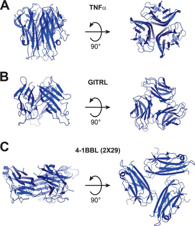
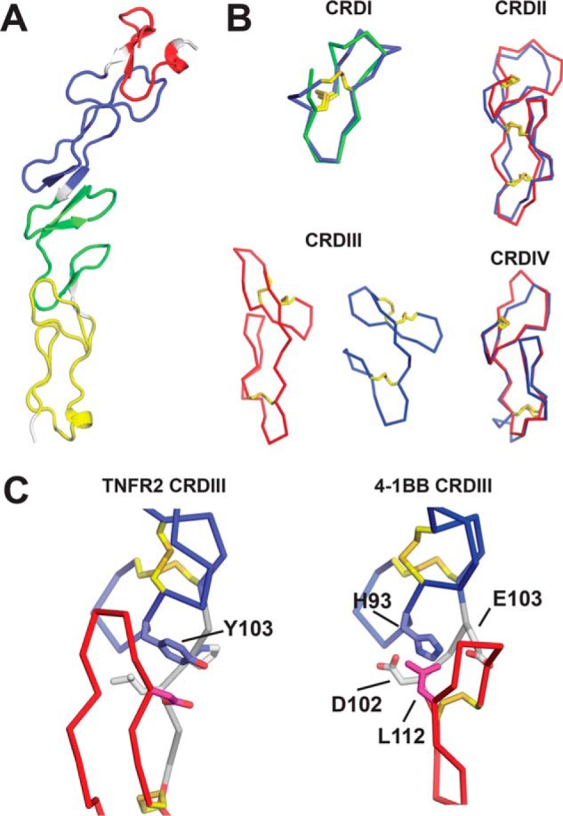
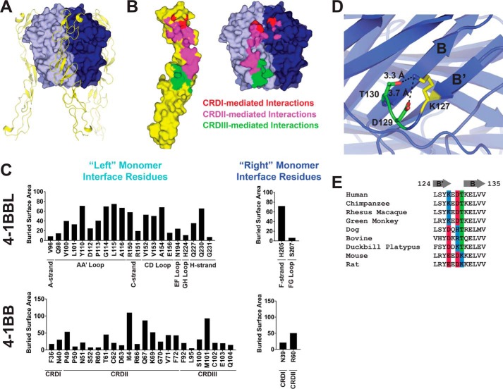
4-1BBL is a member of the tumor necrosis factor (TNF) superfamily and is the ligand for the TNFR superfamily receptor, 4-1BB. 4-1BB plays an immunomodulatory role in T cells and NK cells, and agonists of this receptor have garnered strong attention as potential immunotherapy agents. Broadly speaking, the structural features of TNF superfamily members, their receptors, and ligand-receptor complexes are similar. However, a published crystal structure of human 4-1BBL suggests that it may be unique in this regard, exhibiting a three-bladed propeller-like trimer assembly that is distinctly different from that observed in other family members. This unusual structure also suggests that the human 4-1BB/4-1BBL complex may be structurally unique within the TNF/TNFR superfamily, but to date no structural data have been reported. Here we report the crystal structure of the human 4-1BB/4-1BBL complex at 2.4-Å resolution. In this structure, 4-1BBL does not adopt the unusual trimer assembly previously reported, but instead forms a canonical bell-shaped trimer typical of other TNF superfamily members. The structure of 4-1BB is also largely canonical as is the 4-1BB/4-1BBL complex. Mutational data support the 4-1BBL structure reported here as being biologically relevant, suggesting that the previously reported structure is not. Together, the data presented here offer insight into structure/function relationships in the 4-1BB/4-1BBL system and improve our structural understanding of the TNF/TNFR superfamily more broadly.

Keywords: T-cell biology; crystal structure; protein structure; protein-protein interaction; structural biology; structure-function; tumor necrosis factor (TNF).


Conflict of interest statement

All authors are or have been employees of and may hold financial interests in MedImmune LLC.

Figures

Figure 1.
Figure 1.
Structural features of TNFR superfamily ligands. A, TNFα is representative of the canonical bell-shaped trimer structure observed for most family members (PDB code 3ALQ). B, GITRL is representative of an atypical subgroup of ligands with generally similar structure, but a shorter THD and a more open blooming flower-like trimer conformation (PDB code 2Q1M). C, a published crystal structure of 4-1BBL (e4-1BBL as described by Won et al. (9), PDB ID 2X29) showing a highly unusual three-bladed propeller structure, which is distinct from all other TNFR superfamily ligands.
Figure 2.
Figure 2.
Residues outside the THD of 4-1BBL are not required for function. A, HEK293 cells expressing full-length WT 4-1BBL or variants in which an increasing number of residues upstream of the N-terminal boundary of the TNF homology domain were mutated to Gly/Ser (labeled N1–N5) were incubated with 4-1BB-dependent NF-κB luciferase reporter cells to measure the 4-1BB agonist activity of the ligand constructs. Sequence details for each construct are shown in the schematic. B, same as in A except that 4-1BBL residues 80–88 were fixed as Gly4SerGly4 (N3 variant from A) and an increasing number of residues extending beyond the C-terminal boundary of the THD were deleted in constructs labeled C1–C6. Sequence details for each construct are shown in the schematic. In both experiments 293 cells not expressing 4-1BBL were included as a negative control.
Figure 3.
Figure 3.
THD-only 4-1BBL is a biologically active stable trimer in solution. A, SEC chromatogram and overlaid plot of static light-scattering derived molar mass data for THD-only 4-1BBL. The expected trimer molar mass is indicated with a dotted line. B, SEC chromatogram and overlaid plot of static light-scattering derived molar mass data for the THD-only 4-1BBL/4-1BB complex with components mixed at ∼1:1 molar ratio. The expected molar mass for the 3:3 4-1BB/4-1BBL complex is indicated with a dotted line. C, 4-1BB NF-κB luciferase reporter assay activity for THD-only 4-1BBL. Assay is as described in the legend to Fig. 2 and under “Materials and methods.” Data are shown using reporter cells that are either positive or negative for expression of 4-1BB.
Figure 4.
Figure 4.
The structure of THD-only 4-1BBL. A, overlay of THD-only 4-1BBL monomer (blue) with e4-1BBL monomer from PDB code 2X29 (green). The THD-only 4-1BBL construct is comprised of residues 89–242 of the full-length protein, whereas e4-1BBL is comprised of residues 58–254. The 111–130 residue region, which differs significantly between the two structures, is highlighted for THD-only 4-1BBL (orange) and e4-1BBL (yellow). B, structural comparison of the 111–130 residue region (yellow) in THD-only 4-1BBL (blue, left) and e4-1BBL (green, right). The “X” strand that displaces the A′ and B′ strands in e4-1BBL is highlighted (magenta). C, comparison of the trimer structures of THD-only 4-1BBL (blue) and e4-1BBL (green). D, detailed view of the trimer interface of THD-only 4-1BBL (blue, left) and e4-1BBL (green, right). Tiled aromatic residues that typically pack together to form the trimer core in TNFR superfamily ligands (magenta sticks) are shown as is a linear stretch of C-terminal residues that participates in the e4-1BBL trimer interface (yellow sticks). For each illustration, the monomer on the left-hand side is shown in the same orientation. E, comparison of E-strand contacts in the trimer interfaces of LIGHT (left, red) and THD-only 4-1BBL (right, blue). E-strand residues (yellow) and E-strand contacting residues (cyan) are shown as sticks. F, comparison of GH loop (yellow) conformations in LIGHT (left, red) and THD-only 4-1BBL (right, blue). The AA′ loop, which changes conformation, and the GH loop are indicated as is the side of the monomer involved in the trimer interface.
Figure 5.
Figure 5.
The structure of 4-1BB. A, cartoon of the 4-1BB structure with cysteine-rich domain I (CRDI) (red), CRDII (blue), CRDIII (green), and CRDIV (yellow) indicated. Linker regions connecting the CRDs are colored gray. B, comparison of 4-1BB CRD structures (blue) with those of TNFR2 (red) and TRAILR2 (green). Disulfide bonds (yellow sticks) are also shown. C, comparison of the A2/B1 interface in TNFR2 CRDIII to the A2/A1 interface in 4-1BB CRDIII. In both structures, the N-terminal A2 modules (blue) and downstream B1 or A1 modules (red) are indicated. Linker regions connecting the modules (gray) are also shown as are disulfide bonds (yellow sticks). Key residues involved in the A2/B1 and A2/A1 interfaces are shown and labeled (sticks) and colored to match the module in which they are located.
Figure 6.
Figure 6.
The structure of the 4-1BB/4-1BBL complex. A, structure of the 4-1BB/4-1BBL complex. 4-1BBL is shown in surface representation with neighboring monomer units indicated (blue and cyan). 4-1BB (yellow) is shown as a cartoon. B, open book representation of the 4-1BB/4-1BBL interface (4-1BB has been rotated 180° and translated with respect to 4-1BBL). 4-1BB (left) and 4-1BBL (right) are shown in surface representation with interface residues highlighted. 4-1BB CRDI residues and their contacting partners (red), CRDII residues and their contacting partners (magenta), and CRDIII residues and their contacting partners (green) are indicated. C, buried surface area contributions of individual residues in 4-1BBL (top) and 4-1BB (bottom). Relevant subdomains and secondary structure regions are indicated. Residues involved in the interface with the left and right 4-1BBL monomers as depicted in A and B are segregated in the left and right columns. D, structural detail of the intrachain interactions formed by Lys-127 in 4-1BBL. The B- and B′-strands are labeled. E, sequence alignment of the B′- and B-strand regions of 4-1BBL from diverse species. Positions 127, 129, and 130, which appear to play a role in shaping the local structure of the B′B-strand region in the human 4-1BBL structure reported here, are highlighted and colored according to their side chain charge properties: positive (blue), negative (red), or polar (green).

References

    1. Sanmamed M. F., Pastor F., Rodriguez A., Perez-Gracia J. L., Rodriguez-Ruiz M. E., Jure-Kunkel M., and Melero I. (2015) Agonists of co-stimulation in cancer immunotherapy directed against CD137, OX40, GITR, CD27, CD28, and ICOS. Semin. Oncol. 42, 640–655 10.1053/j.seminoncol.2015.05.014 - DOI - PubMed
    1. Schaer D. A., Hirschhorn-Cymerman D., and Wolchok J. D. (2014) Targeting tumor-necrosis factor receptor pathways for tumor immunotherapy. J. Immunother. Cancer 2, 7 10.1186/2051-1426-2-7 - DOI - PMC - PubMed
    1. Chester C., Sanmamed M. F., Wang J., and Melero I. (2018) Immunotherapy targeting 4-1BB: mechanistic rationale, clinical results, and future strategies. Blood 131, 49–57 - PubMed
    1. Bartkowiak T., and Curran M. A. (2015) 4-1BB agonists: multi-potent potentiators of tumor immunity. Front. Oncol. 5, 117 - PMC - PubMed
    1. Vinay D. S., and Kwon B. S. (2012) Immunotherapy of cancer with 4-1BB. Mol. Cancer Ther. 11, 1062–1070 10.1158/1535-7163.MCT-11-0677 - DOI - PubMed

Publication types