. 2018 May 3;3(9):e98694.
doi: 10.1172/jci.insight.98694.

Phosphodiesterase 2A as a therapeutic target to restore cardiac neurotransmission during sympathetic hyperactivity

Affiliations

Phosphodiesterase 2A as a therapeutic target to restore cardiac neurotransmission during sympathetic hyperactivity

Kun Liu et al. JCI Insight. .

Abstract

Elevated levels of brain natriuretic peptide (BNP) are regarded as an early compensatory response to cardiac myocyte hypertrophy, although exogenously administered BNP shows poor clinical efficacy in heart failure and hypertension. We tested whether phosphodiesterase 2A (PDE2A), which regulates the action of BNP-activated cyclic guanosine monophosphate (cGMP), was directly involved in modulating Ca2+ handling from stellate ganglia (SG) neurons and cardiac norepinephrine (NE) release in rats and humans with an enhanced sympathetic phenotype. SG were also isolated from patients with sympathetic hyperactivity and healthy donor patients. PDE2A activity of the SG was greater in both spontaneously hypertensive rats (SHRs) and patients compared with their respective controls, whereas PDE2A mRNA was only high in SHR SG. BNP significantly reduced the magnitude of the calcium transients and ICaN in normal Wistar Kyoto (WKY) SG neurons, but not in the SHRs. cGMP levels stimulated by BNP were also attenuated in SHR SG neurons. Overexpression of PDE2A in WKY neurons recapitulated the calcium phenotype seen in SHR neurons. Functionally, BNP significantly reduced [3H]-NE release in the WKY rats, but not in the SHRs. Blockade of overexpressed PDE2A with Bay 60-7550 or overexpression of catalytically inactive PDE2A reestablished the modulatory action of BNP in SHR SG neurons. This suggests that PDE2A may be a key target in modulating the action of BNP to reduce sympathetic hyperactivity.

Keywords: Calcium; Cardiology; Neuroscience; Phosphodiesterases.


Conflict of interest statement

Conflict of interest: The authors have declared that no conflict of interest exists.

Figures

Figure 1
Figure 1. Measurements of intracellular cGMP concentration in living cardiac sympathetic neurons in the presence of BNP.
(A) Representative data traces showing dynamics of cytosolic cGMP-induced fluorescence resonance energy transfer (FRET) changes by ratiometric recording of CFP and YFP emission changes in response to increasing concentrations of brain natriuretic peptide (BNP) in spontaneously hypertensive rat (SHR) and Wistar cardiac sympathetic neurons. Saturation of the sensor was achieved using 3-morpholinosydnonimine chloride (SIN-1 chloride; 10 μmol/l; Calbiochem, 16142-27-1) + IBMX (100 μmol/l). (B) Percentage changes in cGMP in response to increasing concentrations of BNP (left, from 10–250 nmol/l) and 100 nmol/l BNP with 1 μmol/l Bay 60-7550 or 100 μmol/l IBMX (right) in SHRs and Wistar rats. *P < 0.05 by unpaired t test. In each case, neurons were derived from 3 or more rats. n indicates number of SG neurons.
Figure 2
Figure 2. Measurement of PDE activity and relative PDE2A mRNA levels in stellate ganglia from human and rat.
(A) Representative data showing cGMP-PDE specific activities and relative PDE2A activities in left and right stellate ganglia tissue from sympathetic hyperactive patients (pathological) and normal control; and (B) in stellate ganglia tissue from 38-week-old Wistar Kyoto (WKY) rats and spontaneously hypertensive rats (SHRs). (C) cAMP-PDE specific activities and relative PDE2A activities in stellate ganglia tissue from 4-week-old WKY rats and SHRs. Quantitative RT-PCR analysis of PDE2A mRNA levels in stellate ganglia tissue of sympathetic hyperactive patients (pathological) and normal control (D) and 4-week-old WKY rats and SHRs (E); data are shown as mean ± SEM. *P < 0.05 by unpaired t test. Left: left stellate ganglion; Right: right stellate ganglion in human. n indicates stellate ganglia number.
Figure 3
Figure 3. Effect of BNP on the norepinephrine release from isolated atria.
(A) Representative group raw data traces showing the time control for [3H]-norepinephrine ([3H]-NE) release from isolated atria harvested from 4-week-old Wistar Kyoto (WKY) rats and spontaneously hypertensive rats (SHRs). The atria were stimulated at 5 Hz for 1 minute at the 16th (S1) and 40th (S2) minutes. (B) Group mean data show no significant changes between S1 and S2 within group in response of 5Hz stimulation evoked [3H]-NE release of time control. However, both S1 and S2 are significantly enhanced in the SHRs compared with the WKY rats (WKY: n = 6, SHR: n = 7). *P < 0.05 by 1-way ANOVA. (C) Same as in A but with the addition of 250 nmol/l brain natriuretic peptide (BNP). Group raw data traces (C) and mean data (D) show that BNP caused a significant decrease in 5-Hz stimulation–evoked [3H]-NE release (S2) in WKY rats, but not in SHRs (WKY: n = 10, SHR: n = 9). *P < 0.05 by 1-way ANOVA. Percentage changes in NE release was expressed as a ratio of increase in NE radioactivity after electrical stimulation over the total radioactivity.
Figure 4
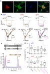
Figure 4. Measurement of calcium current and intracellular free calcium transients in stellate ganglia neurons in the presence of BNP and phosphodiesterase 2A inhibitor.
(A) Fluorescence images of a cultured cardiac sympathetic neurons derived from a 4-week-old Wistar Kyoto (WKY) rat stellate ganglion that was stained for the catecholamine neuronal marker tyrosine hydroxylase (TH, red), phosphodiesterase 2A (PDE2A) antibody (green), costained with the nuclear marker DAPI (blue), and overlay. Scale bar: 20 μm. (B) Representative whole-cell calcium current traces (upper graphs) obtained before and after exposure to 100 nmol/l brain natriuretic peptide (BNP) (left, middle) or 100 nmol/l BNP with 1 μmol/l Bay 60-7550 (right) from 4-week-old WKY rats and spontaneously hypertensive rats (SHRs). Currents were evoked by test pulses of −10 mV from a holding potential of –90 mV. Mean current density–voltage relationships (lower graphs) in the presence and absence of 100 nmol/l BNP (left, middle) or 100 nmol/l BNP with 1 μmol/l Bay 60-7550 (right) from 4-week-old WKY rats and SHRs. *P < 0.05 by paired t test. Vm, membrane voltage in mV; ICa indicates calcium current; pA, picoampere; pF, picofarad. (C) Pharmacological protocols (upper) and raw data trace (lower) recording intracellular calcium transient ([Ca2+]i) in a single cardiac sympathetic neuron from 4-week-old WKY rats and SHRs. A neuron loaded with fura-2 acetoxymethyl ester (Fura-2/AM, 2 μmol/l) was stimulated by 50 mmol/l KCl for 30 seconds to depolarize the neuron and evoke voltage-gated Ca2+ entry. The size of the first (S1) and second (S2) KCl stimulation was compared. (D) Group data showing KCl-evoked peak [Ca2+]i changes expressed as a ratio (%) of S2 compared with S1 in response to 100 or 250 nmol/l BNP with or without PDE2 inhibitor Bay 60-7550 (1 μmol/l) in the WKY rats (upper) and SHRs (lower). *P < 0.05, **P < 0.01, ***P < 0.001, compared with control; P < 0.05, †††P < 0.001; one-way ANOVA. n indicates the number of neurons.
Figure 5
Figure 5. Overexpression of PDE2A in stellate neurons from normotensive rats causes loss of BNP efficacy.
(A) Representative Western blot showing PDE2A.mCherry expression (127 kDa) in Wistar Kyoto (WKY) rat stellate ganglia tissue (with anti-PDE2A antibody) 3 days after transduction with Ad.mCherry virus or Ad.mCherry-PDE2A. Band optical density was normalized to that of β-actin (42 kDa) as a loading control. n = 4 in each group, P < 0.05. (B) Representative calcium current traces (upper graph) and mean current density–voltage relations (lower graph) obtained before and after exposure to 100 nmol/l brain natriuretic peptide (BNP) (a, b) or BNP with 1 μmol/l Bay 60-7550 (c) in the transduced with Ad.mCherry virus or Ad.PDE2A from cardiac sympathetic neurons. *P < 0.05, paired t test. Vm membrane voltage in mV; ICa indicates calcium current; pA, picoampere; pF, picofarad. (C) Percentage change in the peak of intracellular calcium transient ([Ca2+]i) in response of 50 mmol/l KCl for 30 seconds in cardiac sympathetic neurons from WKY rats gene transferred with Ad.mCherry virus or Ad.PDE2A virus in the presence of 100 or 250 nmol/l BNP with or without PDE2A inhibitor Bay 60-7550 (1 μmol/l). *P < 0.05, **P < 0.01, ***P < 0.001, compared with control; P < 0.05; one-way ANOVA. n indicates the number of neurons.
Figure 6
Figure 6. Overexpression of dnPDE2A in stellate neurons from hypertensive rats restores the inhibitory BNP action on calcium signaling.
(A) cGMP-PDE specific total activities in stellate ganglia (SG) tissue from 4-week-old spontaneously hypertensive rats (SHRs) transduced with Ad.mCherry or Ad.dnPDE2A. (B) Representative calcium current traces (upper graphs) and mean current density–voltage relations (lower graphs) obtained before and after exposure to 100 nmol/l BNP in the Ad.mCherry-transduced (left) and Ad.dnPDE2A-transduced (right) SG neurons. *P < 0.05, paired t test. Vm membrane voltage in mV; ICa indicates calcium current; pA, picoampere; pF, picofarad. (C) Percentage change in the peak of intracellular calcium transient ([Ca2+]i) in cardiac sympathetic neurons from SHRs gene transferred with Ad.mCherry (left) or Ad.dnPDE2A (right) virus in the presence of 100 or 250 nmol/l brain natriuretic peptide (BNP) with or without PDE2A inhibitor Bay 60-7550 (1 μmol/l). Both 100 and 250 nmol/l BNP failed to decrease [Ca2+]i with Ad.mCherry virus, unless in the presence of Bay 60-7550 (left). However, 100 and 250 nmol/l BNP decrease [Ca2+]i after transduction with Ad.dnPDE2A (right). *P < 0.05, **P < 0.01, ***P < 0.001, compared with control; P < 0.05, †††P < 0.001; one-way ANOVA. n indicates the number of neurons.
Figure 7
Figure 7. Overexpression of dnPDE2A in atria from hypertensive rat restores the action of BNP on norepinephrine release.
(A) Representative raw data trace showing the effect of 250 nmol/l BNP on [3H]-norepinephrine ([3H]-NE) release during 5-Hz field stimulation from isolated 4-week-old spontaneously hypertensive rat (SHR) atria gene transferred with Ad.mCherry (left) or Ad.dnPDE2A (right) virus. (B) Group mean data show percentage changes in NE release expressed as a ratio of increase in NE radioactivity after 5-Hz field stimulation over the total radioactivity. Brain natriuretic peptide (BNP) (250 nmol/l) significantly reduced [3H]-NE release in SHRs transduced with Ad.dnPDE2A (n = 10), but not in the Ad.mCherry group (n = 8). **P < 0.01, ***P < 0.001 by 1-way ANOVA.
Figure 8
Figure 8. Schematic representation depicting the effect of PDE2A on the regulation of BNP in cardiac neurotransmission in normal and diseased neurons.
(A) Brain natriuretic peptide (BNP) can decrease neurotransmission in healthy neurons by activating the particulate guanylyl cyclase–coupled (pGC-coupled) cGMP pathway to decrease intracellular calcium transients and exocytosis resulting in a decrease in the heart rate (HR) response to sympathetic nerve stimulation. Overexpression of PDE2A in healthy neurons mimics the lack of BNP efficacy seen in diseased neurons. (B) In the diseased neuron, PDE2A is increased and increases the hydrolysis of cGMP and decreases the sympatholytic action of BNP that results in an increase in neurotransmission and HR responses to sympathetic activation. Blockade of overexpressed PDE2A with Bay 60-7550 or overexpression of catalytically inactive PDE2A reestablished the modulatory action of BNP in the diseased neuron. β1-AR, β1-adrenergic receptor; NE, norepinephrine; NPR, natriuretic peptide receptor; SNS, sympathetic nerve stimulation.

References

    1. Tsutamoto T, et al. Attenuation of compensation of endogenous cardiac natriuretic peptide system in chronic heart failure: prognostic role of plasma brain natriuretic peptide concentration in patients with chronic symptomatic left ventricular dysfunction. Circulation. 1997;96(2):509–516. doi: 10.1161/01.CIR.96.2.509. - DOI - PubMed
    1. Richards AM, Crozier IG, Holmes SJ, Espiner EA, Yandle TG, Frampton C. Brain natriuretic peptide: natriuretic and endocrine effects in essential hypertension. J Hypertens. 1993;11(2):163–170. doi: 10.1097/00004872-199302000-00008. - DOI - PubMed
    1. La Villa G, et al. Cardiovascular effects of brain natriuretic peptide in essential hypertension. Hypertension. 1995;25(5):1053–1057. doi: 10.1161/01.HYP.25.5.1053. - DOI - PubMed
    1. Mehel H, et al. Phosphodiesterase-2 is up-regulated in human failing hearts and blunts β-adrenergic responses in cardiomyocytes. J Am Coll Cardiol. 2013;62(17):1596–1606. doi: 10.1016/j.jacc.2013.05.057. - DOI - PubMed
    1. Zaccolo M, Movsesian MA. cAMP and cGMP signaling cross-talk: role of phosphodiesterases and implications for cardiac pathophysiology. Circ Res. 2007;100(11):1569–1578. doi: 10.1161/CIRCRESAHA.106.144501. - DOI - PubMed

Publication types

MeSH terms