Clinical Trial
. 2018 May 3;3(9):e120608.
doi: 10.1172/jci.insight.120608.

First-in-human topical microbiome transplantation with Roseomonas mucosa for atopic dermatitis

Affiliations
Clinical Trial

First-in-human topical microbiome transplantation with Roseomonas mucosa for atopic dermatitis

Ian A Myles et al. JCI Insight. .

Abstract

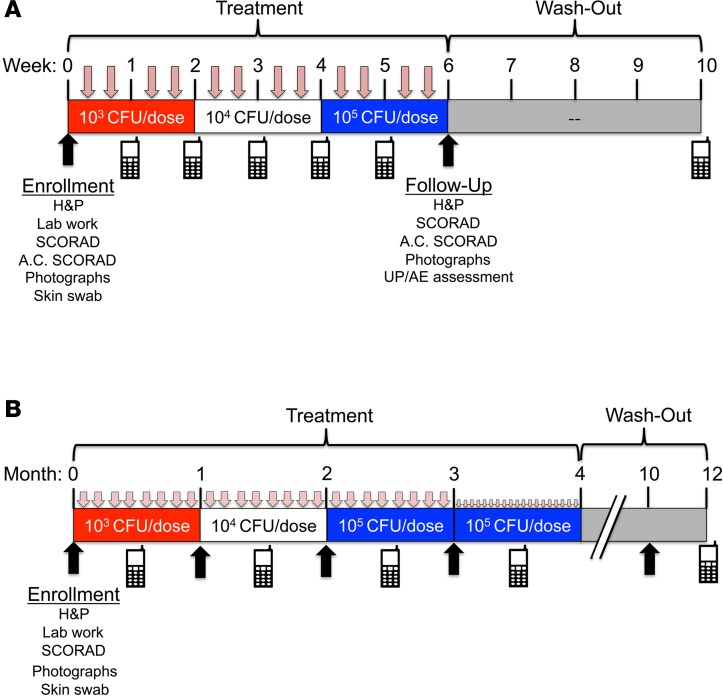
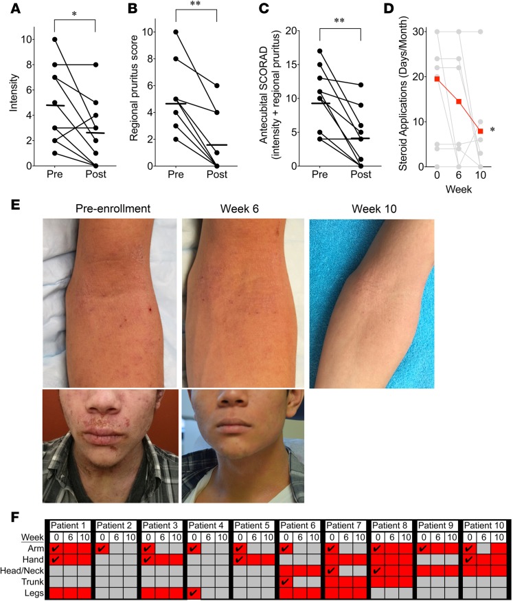
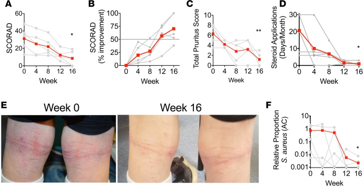
The underlying pathology of atopic dermatitis (AD) includes impaired skin barrier function, susceptibility to Staphylococcus aureus skin infection, immune dysregulation, and cutaneous dysbiosis. Our recent investigation into the potential role of Gram-negative skin bacteria in AD revealed that isolates of one particular commensal, Roseomonas mucosa, collected from healthy volunteers (HVs) improved outcomes in mouse and cell culture models of AD. In contrast, isolates of R. mucosa from patients with AD worsened outcomes in these models. These preclinical results suggested that interventions targeting the microbiome could provide therapeutic benefit for patients with AD. As a first test of this hypothesis in humans, 10 adult and 5 pediatric patients were enrolled in an open-label phase I/II safety and activity trial (the Beginning Assessment of Cutaneous Treatment Efficacy for Roseomonas in Atopic Dermatitis trial; BACTERiAD I/II). Treatment with R. mucosa was associated with significant decreases in measures of disease severity, topical steroid requirement, and S. aureus burden. There were no adverse events or treatment complications. We additionally evaluated differentiating bacterial metabolites and topical exposures that may contribute to the skin dysbiosis associated with AD and/or influence future microbiome-based treatments. These early results support continued evaluation of R. mucosa therapy with a placebo-controlled trial.

Keywords: Allergy; Dermatology; Immunology; Skin.


Conflict of interest statement

Conflict of interest: The authors have declared that no conflict of interest exists.

Figures

Figure 1
Figure 1. Overview of study design.
Summary of study design for adult (A) and pediatric (B) cohorts. Upon enrollment patients underwent history and physical (H&P), screening blood work, bacterial antecubital skin swab, and assessment of antecubital-specific (A.C.) and total SCORAD values. Patients self-administered the topical, live bacteria in a predetermined dose escalation from 103–105 colony-forming units (CFU) per site. Treatments were twice per week (as indicated by pink arrows) for all adults and for weeks 0–12 for the pediatric cohort. Pediatric patients administered treatments every other day during weeks 13–16. Treatments consisted only of R. mucosa in 250 μl of 10%–15% sucrose. Remote follow-up for solicitation of unexpected problems and/or adverse events (UP/AE; indicated by cell phone icons) was performed weekly during adult treatment and then 4 weeks after discontinuation of therapy. Washout evaluation for pediatric cohort is ongoing.
Figure 2
Figure 2. Topical Roseomonas mucosa shows activity against atopic dermatitis in adults.
Mean (bars) and individual (circles; n = 10) before- and after-treatment scores for objective intensity (A) and subjective pruritus (B) as measured by SCORAD. (C) Antecubital-specific SCORAD; sum of local intensity and pruritus scores. (D) Mean (scarlet) and individual (gray) self-reported steroid use (days/month) from the 6 weeks prior to enrollment (week 0), after treatment (week 6), and after washout (week 10). Patients were instructed to maintain their home regimens throughout active treatment; however, patients 2 and 9 discontinued topical steroids upon initiation of R. mucosa treatment. (E) Pretreatment photos from enrollment, after treatment, and after washout (photo taken by patient) for the dominant-arm antecubital fossa and the face for patient 7 (see Supplemental Table 2). (F) Summary of areas of involvement (scarlet shading), asymptomatic areas (gray), and areas directly treated (check mark). Significance determined by 2-tailed Student’s t test and nonparametric Wilcoxon’s matched-pairs test. *P < 0.05, **P < 0.01.
Figure 3
Figure 3. Topical Roseomonas mucosa shows activity against atopic dermatitis in children.
Mean (scarlet) and individual (gray; n = 5) SCORAD values (A) and percentage improvement (B) during treatment. Dotted line (B) indicates improvement level that is inconsistent with null hypothesis (see Methods). (C) Mean and individual pruritus. (D) Mean and individual patient-reported days of topical steroid use per month for the 3 months prior to enrollment (week 0) and during treatment. (E) Pretreatment photos from enrollment (week 0), after treatment (week 16) for the popliteal fossae for patient S5 (see Supplemental Table 1). (F) Ratio of Staphylococcus aureus to coagulase-negative staphylococci from the antecubital (AC) fossa as determined by culture. Significance determined by 2-tailed Student’s t test and nonparametric Wilcoxon’s matched-pairs test. *P < 0.05, **P < 0.01 as determined versus enrollment value.
Figure 4
Figure 4. Strains of Roseomonas mucosa are impacted by environmental exposures.
(A) Representative pictures for mean zone of inhibition (ZOI) on agar growth plates for R. mucosa from healthy volunteers (HVs), R. mucosa from patients with atopic dermatitis (AD), or Staphylococcus aureus. (B) Mean (bar) and individual (dots) ZOI for uncommon (B) and common (C) topical exposures. (D) Mean and individual ZOI for individual paraben (-pb) subtypes. (E) As in A, imaging of resistant S. aureus colonies in response to methyl-, ethyl-, propyl-, and benzyl-paraben exposure. (F) Mean and individual ZOI for common topical emollients. (G) Mean and individual ZOI for dilute bleach. Data are representative of 3 independent experiments using n = 3–7 different isolates per group, per experiment and displayed as mean ± SEM. *P < 0.05, **P < 0.01, ***P < 0.001, ****P < 0.0001 as determined by ANOVA, significance indicated as compared with R. mucosa (HV) group.

Similar articles

Cited by

References

    1. Eichenfield LF, Ahluwalia J, Waldman A, Borok J, Udkoff J, Boguniewicz M. Current guidelines for the evaluation and management of atopic dermatitis: A comparison of the Joint Task Force Practice Parameter and American Academy of Dermatology guidelines. J Allergy Clin Immunol. 2017;139(4S):S49–S57. - PubMed
    1. Bantz SK, Zhu Z, Zheng T. The atopic march: progression from atopic dermatitis to allergic rhinitis and asthma. J Clin Cell Immunol. 2014;5:202. - PMC - PubMed
    1. Boguniewicz M, Leung DY. Recent insights into atopic dermatitis and implications for management of infectious complications. J Allergy Clin Immunol. 2010;125(1):4–13. - PMC - PubMed
    1. Latvala J, von Hertzen L, Lindholm H, Haahtela T. Trends in prevalence of asthma and allergy in Finnish young men: nationwide study, 1966-2003. BMJ. 2005;330(7501):1186–1187. doi: 10.1136/bmj.38448.603924.AE. - DOI - PMC - PubMed
    1. Silverberg JI. Public health burden and epidemiology of atopic dermatitis. Dermatol Clin. 2017;35(3):283–289. doi: 10.1016/j.det.2017.02.002. - DOI - PubMed

Publication types

MeSH terms