. 2018 May 3;3(9):e99357.
doi: 10.1172/jci.insight.99357.

Efficient exon skipping of SGCG mutations mediated by phosphorodiamidate morpholino oligomers

Affiliations

Efficient exon skipping of SGCG mutations mediated by phosphorodiamidate morpholino oligomers

Eugene J Wyatt et al. JCI Insight. .

Abstract

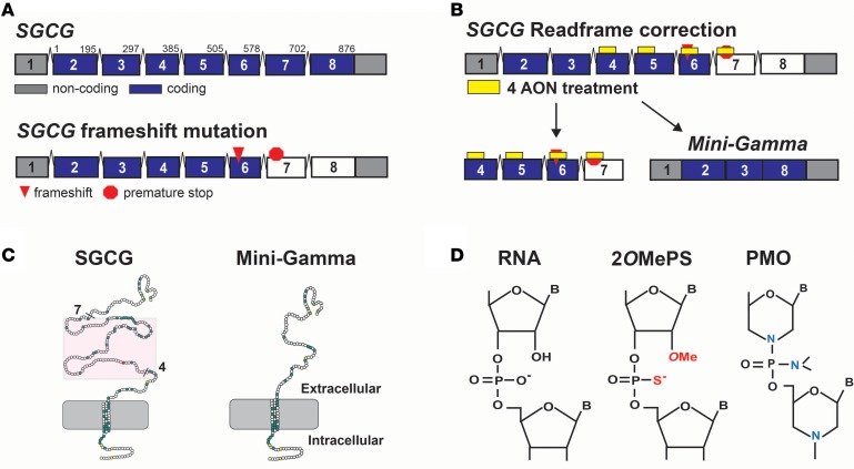
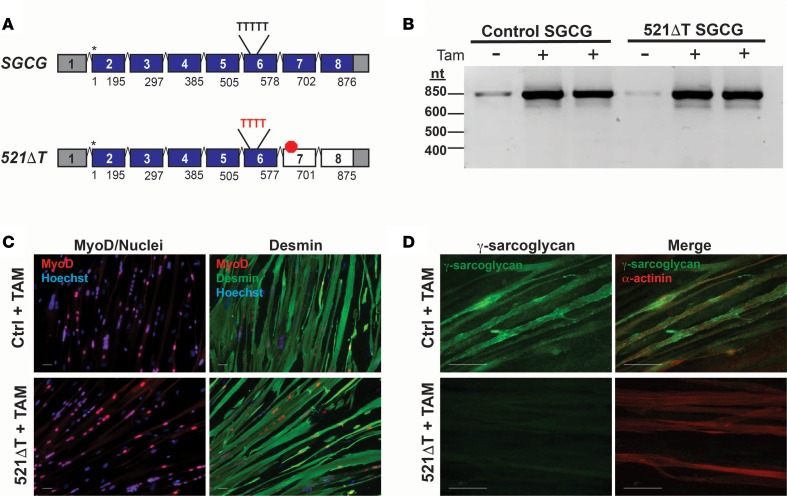
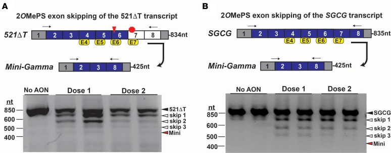
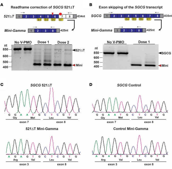
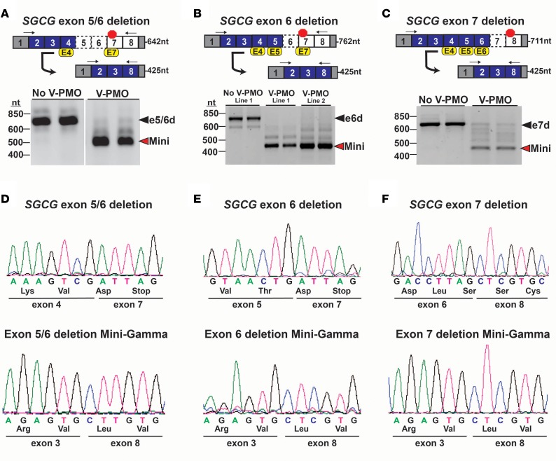
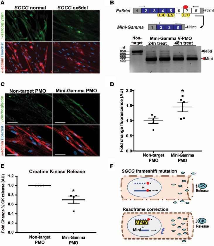
Exon skipping uses chemically modified antisense oligonucleotides to modulate RNA splicing. Therapeutically, exon skipping can bypass mutations and restore reading frame disruption by generating internally truncated, functional proteins to rescue the loss of native gene expression. Limb-girdle muscular dystrophy type 2C is caused by autosomal recessive mutations in the SGCG gene, which encodes the dystrophin-associated protein γ-sarcoglycan. The most common SGCG mutations disrupt the transcript reading frame abrogating γ-sarcoglycan protein expression. In order to treat most SGCG gene mutations, it is necessary to skip 4 exons in order to restore the SGCG transcript reading frame, creating an internally truncated protein referred to as Mini-Gamma. Using direct reprogramming of human cells with MyoD, myogenic cells were tested with 2 antisense oligonucleotide chemistries, 2'-O-methyl phosphorothioate oligonucleotides and vivo-phosphorodiamidate morpholino oligomers, to induce exon skipping. Treatment with vivo-phosphorodiamidate morpholino oligomers demonstrated efficient skipping of the targeted exons and corrected the mutant reading frame, resulting in the expression of a functional Mini-Gamma protein. Antisense-induced exon skipping of SGCG occurred in normal cells and those with multiple distinct SGCG mutations, including the most common 521ΔT mutation. These findings demonstrate a multiexon-skipping strategy applicable to the majority of limb-girdle muscular dystrophy 2C patients.

Keywords: Genetics; Monogenic diseases; Muscle Biology; Neuromuscular disease; Skeletal muscle.


Conflict of interest statement

Conflict of interest: EMM and EJW are coinventors of a patent related to exon skipping (US patent 9,777,271).

Figures

Figure 1
Figure 1. An exon-skipping strategy to treat LGMD 2C.
(A) The SGCG gene, which encodes γ-sarcoglycan, is comprised of 8 exons, and many SGCG mutations disrupt the transcript reading frame, causing Limb-girdle muscular dystrophy (LGMD) 2C. (B) Correction of the reading frame requires the skipping of SGCG exons 4, 5, 6, and 7 at the pre-mRNA level. Four antisense oligonucleotides (AON) were designed to exclude these exons from the mature mRNA transcript. The bypass of exons 4–7 generates an internally truncated product termed Mini-Gamma encoded by exons 2, 3, and 8. (C) Mini-Gamma protein lacks a portion of the extracellular domain encoded by exons 4–7 (red box) and retains the essential cytosolic, transmembrane, and extracellular domains required for functionality (30). (D) Exon skipping AONs are chemically modified to avoid nuclease degradation and destruction of the mRNA transcript. Shown are the chemical structures of 2 types of AONs used for exon skipping. 2’-O-methyl phosphorothioates (2OMePS) include a methyl group on the ribose ring and substitution of sulfur for oxygen to create a phosphothioate bond (red). Phosphorodiamidate morpholino oligomers (PMOs) incorporate morpholino rings rather than ribose rings and substitute nitrogen for oxygen to create a phosphorodiamidate bond (blue) (18). Vivo-PMOs are synthesized with a covalently linked 3′ octa-guanidine delivery moiety for enhanced cell delivery (31).
Figure 2
Figure 2. SGCG mRNA and protein expression in fibroblasts reprogrammed into the myogenic lineage.
(A) Schematic of the SGCG pre-mRNA transcript. The most common LGMD 2C mutation is a single thymine (T) deletion from a string of 5Ts in SGCG exon 6. This mutation, designated 521ΔT, results in a transcript frame shift, which generates a premature stop codon (red octagon). Asterisks represent the transcription start sites, and the numbers denote the mRNA region encoded by each exon. Fibroblasts from a normal control subject (SGCG intact) and a LGMD 2C individual with the 521ΔT mutation were directly reprogrammed into the myogenic lineage using a tamoxifen (Tam) inducible MyoD lentiviral construct (iMyoD) (32). (B) RT-PCR analysis demonstrated expression of the SGCG mRNA in normal control and 521ΔT cells after tamoxifen induction and culture in differentiation media (5 μM 4OH-tamoxifen, 48 hours, 9 days differentiation). (C) Immunofluorescence microscopy (IFM) demonstrated the efficient transduction and myogenic reprogramming of both the control and 521ΔT cell lines (5 μM 4OH-tamoxifen, 48 hours, 12 days differentiation). Nuclear expression of MyoD (red) is observed in the iMyoD-transduced cells. Reprogrammed cells expressed the myogenic marker desmin (green) and formed multinucleated myotubes. Nuclei were labeled with Hoechst 3342 (blue). Scale bar: 50 μM. (D) γ-Sarcoglycan protein (green) was readily detected in the reprogrammed control cells, but not in reprogrammed 521ΔT cells, in accordance with their mutation status. Expression of α-actinin (red) indicated the myogenic reprogramming of each cell line. Scale bar: 50 μM.
Figure 3
Figure 3. 2OMePS-mediated exon skipping of the SGCG transcript.
(A) Correction of the SGCG 521ΔT frameshift mutation (red triangle) requires a multi-AON exon-skipping strategy targeting exons 4, 5, 6, and 7 (yellow boxes) to generate the reading frame–corrected Mini-Gamma transcript encoded by exons 2, 3, and 8. Arrows denote the location of the RT-PCR primers, and the expected amplicon lengths are indicated. Myogenic cells were exposed to a multiexon–skipping cocktail of 2OMePS AONs targeting exons 4, 5, 6, and 7. RT-PCR analysis demonstrated the expression of the 521ΔT transcript (black arrowhead), along with PCR products representing the skipping of 1, 2, and 3 exons (white arrowheads). A PCR product representing the 4-AON–mediated generation of the Mini-Gamma was observed at the expected size (red arrowhead). (B) Treatment of reprogrammed normal control fibroblasts with 2OMePS AONs targeting exons 4, 5, 6, and 7 also induced exon skipping. RT-PCR analysis demonstrated the expression of the SGCG transcript (black arrowhead), along with PCR products representing the skipping of 1, 2, and 3 exons (white arrowheads). No PCR product representing the 4-AON–mediated generation of the Mini-Gamma was observed at the expected size (red arrowhead).
Figure 4
Figure 4. Vivo-PMO–mediated exon skipping of the SGCG transcript.
(A) A multi-AON exon-skipping cocktail using vivo-PMO chemistry was employed to correct the SGCG 521ΔT reading frame in myogenic cells. RT-PCR analysis showed treatment with vivo-PMOs targeting exons 4, 5, 6, and 7 generated the Mini-Gamma reading frame–corrected transcript (red arrowhead) with minimal residual expression of the mutant 521ΔT transcript (black arrowhead). Arrows in schematic denote the location of the RT-PCR primers, and the expected amplicon lengths are indicated. (B) Normal control myogenic cells were treated with the same multiexon-skipping vivo-PMO cocktail. RT-PCR analysis showed expression of the Mini-Gamma transcript (red arrowhead), indicating that normal SGCG transcript could be skipped using vivo-PMOs. Expression of the SGCG transcript in untreated cells is indicated (black arrowhead). (C and D) Sequence analysis of PCR products confirmed the generation of the Mini-Gamma transcripts in each of the mutant cell lines, with the splicing of SGCG exons 3 and 8 demonstrating the removal of exons 4, 5, 6, and 7 from the mature transcripts.
Figure 5
Figure 5. Reading frame correction of multiple SGCG frameshift mutations.
(A–C) Top: Mutation-specific multiexon-skipping strategies were designed to correct the SGCG reading frame of multiple unrelated LGMD 2C patients whose primary SGCG mutations include (A) the deletion of exons 5 and 6; (B) the deletion of exon 6; and (C) the deletion of exon 7. Myogenic cells were treated with vivo-PMO exon skipping cocktails. Arrows denote the location of the RT-PCR primers, and the expected amplicon lengths are indicated. (A–C) Bottom: RT-PCR analysis confirmed the mutant SGCG transcripts were expressed in the reprogrammed cells at the expected size, consistent with their exon deletion status (black arrowheads). Cells treated with the mutation-specific vivo-PMOs cocktails demonstrated predominate expression of the reading frame–corrected Mini-Gamma transcript (red arrowheads). (D–F) Sequence analysis of RT-PCR products: (D) a deletion of exons 5 and 6; (E) a deletion of exon 6; and (F) a deletion of exon 7. Sequence analysis also confirmed the generation of the Mini-Gamma transcript in each of the mutant cell lines, with the splicing of SGCG exons 3 and 8 demonstrating the removal of (D) exons 4 and 7; (E) exon 4, 5, and 7; and (F) exons 4, 5 and 6 from the mature transcripts.
Figure 6
Figure 6. Restoration of SGCG expression as Mini-Gamma protein.
Reprogrammed fibroblasts were treated for 3 days with either a nontargeting control vivo-PMO (nontarget) or a mutation-specific vivo-PMO exon skipping cocktail (Mini-Gamma). (A) Representative IFM images of reprogrammed 521ΔT cells that were treated as indicated. Treatment with reading frame–correcting vivo-PMOs resulted in the expression of the Mini-Gamma protein (green). Myotubes were labeled with α-actinin (red), and nuclei were labeled with Hoechst 3342 (blue). (B) Image analysis demonstrated a significant increase in Mini-Gamma fluorescence after treatment with vivo-PMOs that corrected the transcript reading frame (n = 6) as compared with nontargeting controls (n = 5). (C and D) Reprogrammed cells harboring a deletion of SGCG exons 5 and 6 (ex5/6del) were treated as indicated. IFM image analysis demonstrated a significant increase in Mini-Gamma fluorescence after treatment with vivo-PMOs that corrected the reading transcript reading frame (n = 4) as compared with nontargeting controls (n = 3). (E and F) Reprogrammed cells harboring a deletion of SGCG exons 6 (ex6del) were treated as indicated. IFM image analysis demonstrated a significant increase in Mini-Gamma fluorescence after treatment with vivo-PMOs that corrected the transcript reading frame (n = 5) as compared with nontargeting controls (n = 3). Data represent the mean γ-sarcoglycan fluorescence per α-actinin–positive area normalized to the mean of the untreated group. A minimum of 3 independent fields were analyzed for each sample. *P < 0.05 as determined by 2-tailed Student’s t test. Data represent the mean ± SEM. Scale bars: 50 μM.
Figure 7
Figure 7. SGCG reading frame correction in urine-derived cells (UDCs).
UDCs from a normal control subject and an LGMD 2C patient with a deletion of exon 6 (ex6del) were reprogrammed into a myogenic lineage. (A) γ-Sarcoglycan protein (green) was detected by IFM in the reprogrammed normal control myotubes but not in reprogrammed ex6del cells. α-Actinin, red; nuclei, blue. (B) RT-PCR analysis demonstrated reading frame–corrected Mini-Gamma transcript expression (red arrowhead). (C) Representative IFM images showed the restoration of γ-sarcoglycan protein expression in cells treated with Mini-Gamma vivo-PMOs. (D) Significant increase in γ-sarcoglycan protein fluorescence was observed after treatment with vivo-PMOs (n = 5) as compared with nontargeting control vivo-PMOs (n = 5). A minimum of 3 independent fields were analyzed for each sample. (E) To assess membrane stability in response to vivo-PMO treatment, reprogrammed cells were challenged with hypo-osmotic shock and membrane leak was monitored by release of creatine kinase (CK). Vivo-PMO treatment significantly decreased the relative amount of CK release consistent with increased membrane stability. Data represent the percent of CK released relative to the total CK from 4 independent experiments (n = 3–4, for each). Data are presented as the mean CK released in cells treated with exon-skipping vivo-PMOs relative to the mean in cells treated with a nontargeting vivo-PMO. (F) Model depicting the increased membrane stability that resulted from vivo-PMO–mediated reading frame correction of an SGCG frameshift mutation. *P < 0.05 as determined by 2-tailed Student’s t test. Data represent the mean ± SEM. Scale bars: 50 μM.

References

    1. Fayssoil A, et al. Natural History of Cardiac and Respiratory Involvement, Prognosis and Predictive Factors for Long-Term Survival in Adult Patients with Limb Girdle Muscular Dystrophies Type 2C and 2D. PLoS ONE. 2016;11(4):e0153095. doi: 10.1371/journal.pone.0153095. - DOI - PMC - PubMed
    1. Kirschner J, Lochmüller H. Sarcoglycanopathies. Handb Clin Neurol. 2011;101:41–46. doi: 10.1016/B978-0-08-045031-5.00003-7. - DOI - PubMed
    1. Noguchi S, et al. Mutations in the dystrophin-associated protein gamma-sarcoglycan in chromosome 13 muscular dystrophy. Science. 1995;270(5237):819–822. doi: 10.1126/science.270.5237.819. - DOI - PubMed
    1. Ervasti JM, Campbell KP. Membrane organization of the dystrophin-glycoprotein complex. Cell. 1991;66(6):1121–1131. doi: 10.1016/0092-8674(91)90035-W. - DOI - PubMed
    1. Ozawa E, Mizuno Y, Hagiwara Y, Sasaoka T, Yoshida M. Molecular and cell biology of the sarcoglycan complex. Muscle Nerve. 2005;32(5):563–576. doi: 10.1002/mus.20349. - DOI - PubMed

Publication types

Supplementary concepts