. 2018 May 2;8(1):6865.
doi: 10.1038/s41598-018-25130-3.

The stage of seed development influences iron bioavailability in pea (Pisum sativum L.)

Affiliations

The stage of seed development influences iron bioavailability in pea (Pisum sativum L.)

Katie L Moore et al. Sci Rep. .

Abstract

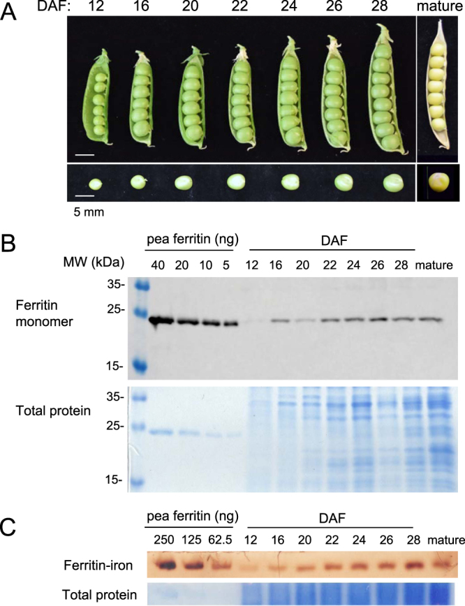
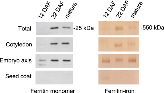
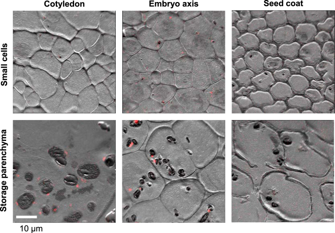
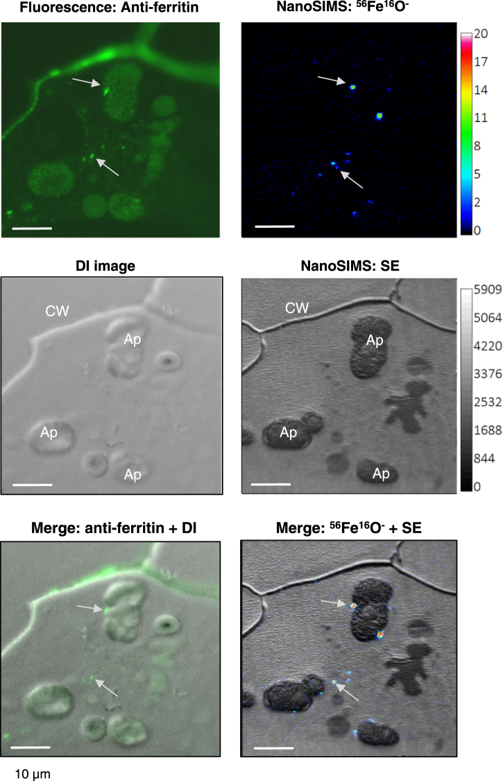
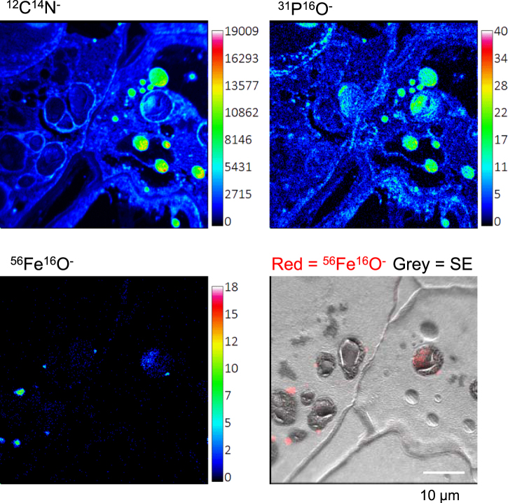
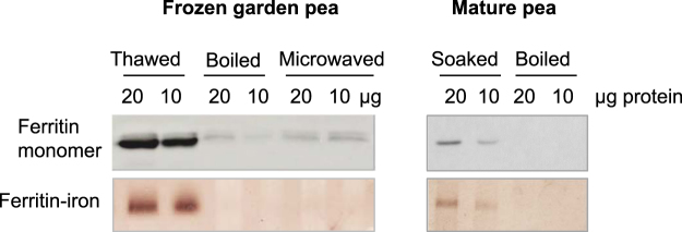
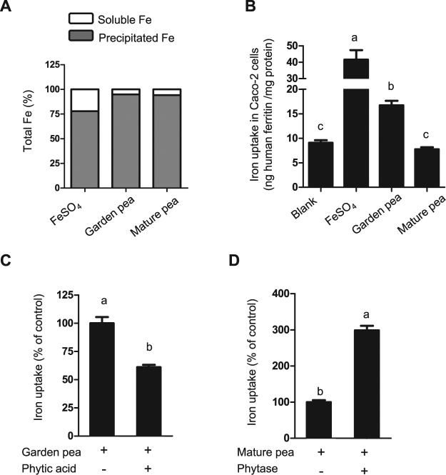
Pea seeds are widely consumed in their immature form, known as garden peas and petit pois, mostly after preservation by freezing or canning. Mature dry peas are rich in iron in the form of ferritin, but little is known about the content, form or bioavailability of iron in immature stages of seed development. Using specific antibodies and in-gel iron staining, we show that ferritin loaded with iron accumulated gradually during seed development. Immunolocalization and high-resolution secondary ion mass spectrometry (NanoSIMS) revealed that iron-loaded ferritin was located at the surface of starch-containing plastids. Standard cooking procedures destabilized monomeric ferritin and the iron-loaded form. Iron uptake studies using Caco-2 cells showed that the iron in microwaved immature peas was more bioavailable than in boiled mature peas, despite similar levels of soluble iron in the digestates. By manipulating the levels of phytic acid in the digestates we demonstrate that phytic acid is the main inhibitor of iron uptake from mature peas in vitro. Taken together, our data show that immature peas and mature dry peas contain similar levels of ferritin-iron, which is destabilized during cooking. However, iron from immature peas is more bioavailable because of lower phytic acid levels compared to mature peas.


Conflict of interest statement

The authors declare no competing interests.

Figures

Figure 1
Figure 1
Ferritin protein and iron loading during development of pea seeds. (A) Images of opened seed pods at the indicated days after flowering (DAF). (B) Protein blot analysis of ferritin in extracts from pea seeds. A dilution series of purified pea ferritin (40 − 5 ng) was run in the four left lanes for comparison with pea extracts (5 µg protein per lane). (C) Iron staining associated with assembled ferritin shells. A dilution series of freshly purified pea ferritin (250–62.5 ng protein, or 55 – 14 ng Fe) and protein extracts from developing peas (20 µg protein per lane) were separated by native gel electrophoresis and stained for iron with Perls’ staining and diaminobenzidine enhancement. The data shown are representative of 4 independent time courses. A second data set is shown in Figure S2.
Figure 2
Figure 2
Ferritin levels in cotyledons, embryo axis and seed coat. Protein extracts (20 µg) from total pea seeds (JI1194), cotyledons, embryo axis and seed coat at three stages of development were separated by SDS-PAGE for immunoblot analysis (left panels) or by native PAGE for iron staining (right panels). The results are representative of 3 independent biological replicates, and each sample was prepared from 2–3 seeds or tissues.
Figure 3
Figure 3
Discrete spots of iron are associated with amyloplasts in pea seeds. NanoSIMS localization of iron in sections of immature pea seeds (JI1194) at 22–24 DAF. The images are representative of 6 analysed regions from the cotyledon, 6 from the embryo axis and 2 regions from the seed coat, showing either small cell types or large storage parenchyma cells. The ion-induced secondary electron image is shown in grey scale and the 56Fe16O signal is shown superimposed in red. Images with the 56Fe16O signal only are shown in Figure S5. The dark oval features observed in the larger cells are starch-filled amyloplasts.
Figure 4
Figure 4
Corresponding localization of iron and ferritin protein. Adjacent thin sections of cotyledons from immature pea seeds (JI1194) at 22–24 DAF were labelled with antibodies against pea ferritin followed by Alexa-Fluor conjugated secondary antibodies (top left, green) or imaged for 56Fe16O ions using NanoSIMS (top right, with colour scale for the intensity of the signal). The differential interference (DI) image of the immunolabelled section and the secondary electron (SE) image for NanoSIMS show the cell walls and organelles. CW, cell wall; Ap, amyloplast. The images are representative of 2 different regions for which 2 cells each were imaged.
Figure 5
Figure 5
Colocalization of Fe with protein and PO compounds. NanoSIMS images of 12C14N (top left), 31P16O (top right), and 56Fe16O (bottom two images) in cotyledons of immature pea seeds (JI1194) at 22–24 DAF. The images show part of neighbouring cells representative of the cotyledons. The image bottom left shows the dense iron spots on the outside of amyloplasts as well as the diffuse pattern of iron inside the amyloplasts (overlay of 56Fe16O in red and secondary electrons, SE, in grey scale). The images are representative of 4 different regions each showing colocalization of Fe with PO compounds.
Figure 6
Figure 6
Ferritin in immature frozen peas and mature dry pea seeds is destabilized by different cooking methods. Frozen garden peas were boiled or microwaved, and mature dry peas were soaked and boiled. Protein extracts were analysed for ferritin monomer by protein blot analysis (top panel) and for iron associated with ferritin by in-gel Perls’/DAB staining (lower panel). The data are representative of 2 biological repeats.
Figure 7
Figure 7
Iron in cooked immature garden peas is more bioavailable than in mature peas. (A) Percentage of soluble and precipitated iron in the final step of the simulated gastrointestinal digestion. (B) Iron uptake in Caco-2 cells exposed to the digestates of microwaved (immature) garden peas and boiled mature peas. (C) Percentage of change in iron uptake into Caco-2 cells after adding phytic acid to the simulated digestion of garden peas, and (D) after adding phytase enzyme to the simulated digestion of mature peas. Data represent means ± SEM from 3–4 different simulated digestions which were each applied in triplicate to Caco-2 cells. Means without a common letter differ (p < 0.05, using one-way ANOVA for (B) and Student t-test in (C and D).

References

    1. Hurrell R, Egli I. Iron bioavailability and dietary reference values. Am. J. Clin. Nutr. 2010;91:1461S–1467S. doi: 10.3945/ajcn.2010.28674F. - DOI - PubMed
    1. Connorton JM, Balk J, Rodríguez-Celma J. Iron homeostasis in plants – a brief overview. Metallomics. 2017;9:813–823. doi: 10.1039/C7MT00136C. - DOI - PMC - PubMed
    1. Theil EC. Iron, ferritin, and nutrition. Annu. Rev. Nutr. 2004;24:327–343. doi: 10.1146/annurev.nutr.24.012003.132212. - DOI - PubMed
    1. Hoppler M, Zeder C, Walczyk T. Quantification of ferritin-bound iron in plant samples by isotope tagging and species-specific isotope dilution mass spectrometry. Anal. Chem. 2009;81:7368–7372. doi: 10.1021/ac900885j. - DOI - PubMed
    1. Briat JF, Duc C, Ravet K, Gaymard F. Ferritins and iron storage in plants. Biochim Biophys Acta. 2010;1800:806–814. doi: 10.1016/j.bbagen.2009.12.003. - DOI - PubMed

Publication types