. 2018 May 2;8(1):6913.
doi: 10.1038/s41598-018-25263-5.

FTY720 Decreases Tumorigenesis in Group 3 Medulloblastoma Patient-Derived Xenografts

Affiliations

FTY720 Decreases Tumorigenesis in Group 3 Medulloblastoma Patient-Derived Xenografts

Evan F Garner et al. Sci Rep. .

Abstract

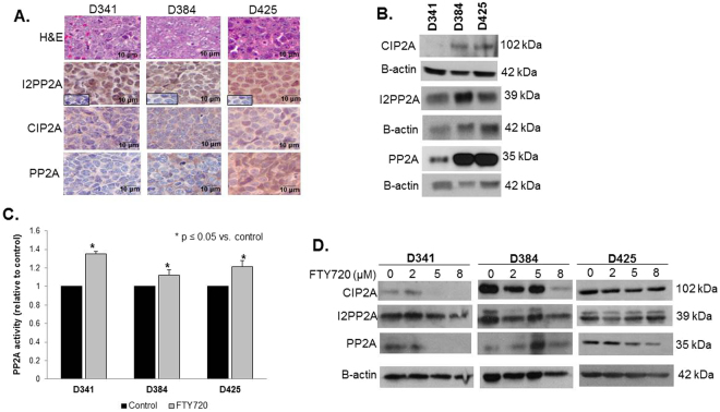
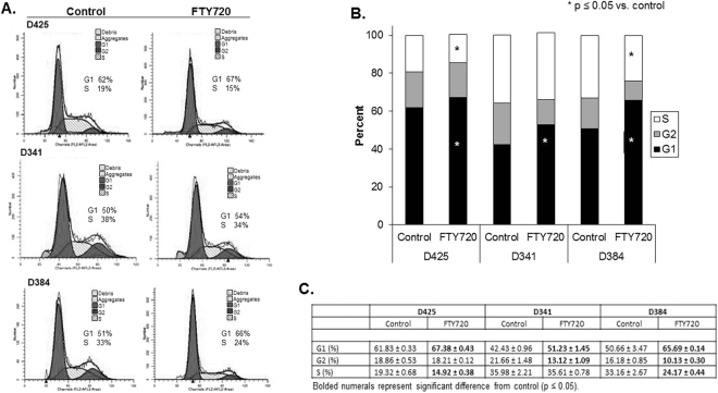
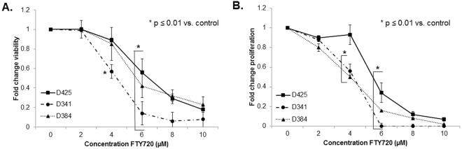
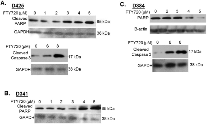
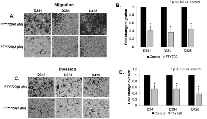
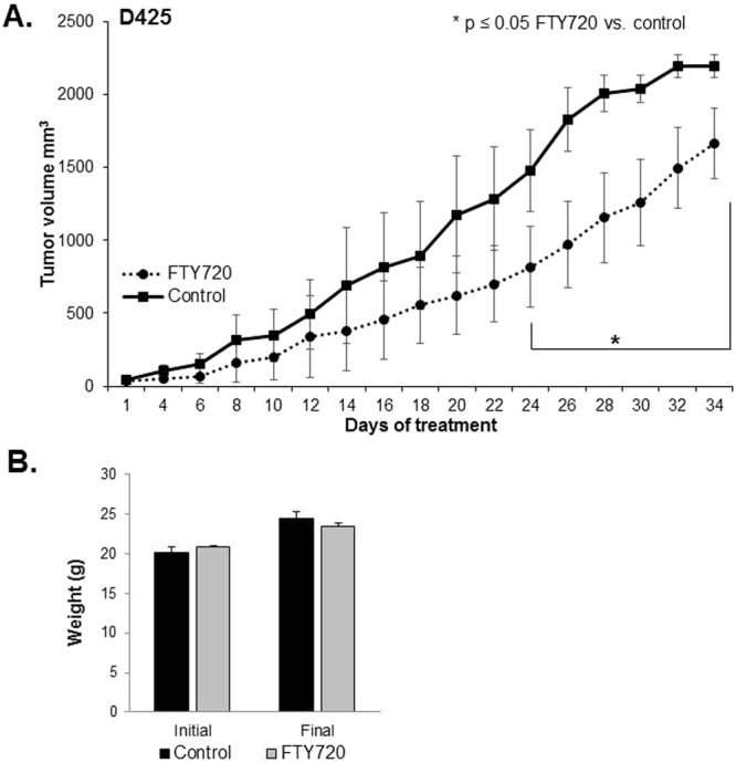
Group 3 tumors account for 28% of medulloblastomas and have the worst prognosis. FTY720, an immunosuppressant currently approved for treatment of multiple sclerosis, has shown antitumor effects in several human cancer cell lines. We hypothesized that treatment with FTY720 (fingolimod) would decrease tumorigenicity in medulloblastoma patient-derived xenografts (PDXs). Three Group 3 medulloblastoma PDXs (D341, D384 and D425) were utilized. Expression of PP2A and its endogenous inhibitors I2PP2A and CIP2A was detected by immunohistochemistry and immunoblotting. PP2A activation was measured via phosphatase activation kit. Cell viability, proliferation, migration and invasion assays were performed after treatment with FTY720. Cell cycle analysis was completed using flow cytometry. A flank model using D425 human medulloblastoma PDX cells was used to assess the in vivo effects of FTY720. FTY720 activated PP2A and led to decreased medulloblastoma PDX cell viability, proliferation, migration and invasion and G1 cell cycle arrest in all three PDXs. FTY720 treatment of mice bearing D425 medulloblastoma PDX tumors resulted in a significant decrease in tumor growth compared to vehicle treated animals. FTY720 decreased viability, proliferation, and motility in Group 3 medulloblastoma PDX cells and significantly decreased tumor growth in vivo. These results suggest that FTY720 should be investigated further as a potential therapeutic agent for medulloblastoma.


Conflict of interest statement

The authors declare no competing interests.

Figures

Figure 1
Figure 1
FTY720 treatment increased protein phosphatase 2A activity. (A) H&E staining confirmed histology consistent with medulloblastoma in PDXs (top panels). Immunohistochemistry was performed to demonstrate I2PP2A, CIP2A and PP2A expression in formalin-fixed, paraffin-embedded human medulloblastoma PDXs. IgG negative controls reacted appropriately (second row panels, lower left insets). (B) Immunoblotting confirmed I2PP2A, CIP2A, and PP2A expression in whole cell lysates from these 3 medulloblastoma PDXs. (C) PP2A activity was measured in all 3 medulloblastoma PDXs. Treatment with FTY720 (5 µM) for 4 hours resulted in a significant increase in PP2A activity in all three PDX’s tested (*p ≤ 0.05). Experiments were repeated at least in triplicate and reported as mean ± SEM. (D) Protein levels of CIP2A and I2PP2A (SET) were measured following FTY720 treatment. CIP2A was diminished in the D341 and D384 MB PDXs (top left and middle panels), but not in the D425 MB PDX (top right panel). I2PP2A levels were not altered (middle panels).
Figure 2
Figure 2
FTY720 treatment of medulloblastoma PDXs led to cell cycle arrest. (A) Representative histograms for cell cycle analysis of D425, D341, and D384 human medulloblastoma PDX cells following treatment with FTY720 (5 µM for 24 hours). Cells were analyzed by flow cytometry following staining with propidium iodine. There was an increase in the percentage of cells in the G1 phase and a decrease in the percentage in S phase following FTY720 treatment. (B) Graphic representation of cell cycle analysis in D425, D341 and D384 medulloblastoma PDX cells treated with FTY720. There was a significant increase in the G1 phase in cells from all three PDXs (*p ≤ 0.05) and a significant decrease in S phase in the D425 and D384 cells (*p ≤ 0.05) after FTY720 treatment, indicating G1 cell cycle arrest. Experiments were repeated at least in triplicate and reported as mean ± SEM. (C) Cell cycle data presented in tabular form reporting mean ± standard error of the mean (SEM). Statistically significant changes are noted with bold type (p ≤ 0.05).
Figure 3
Figure 3
FTY720 treatment resulted in decreased viability and proliferation of medulloblastoma PDXs. (A) Cell viability was measured using alamarBlue® assays. D425, D341 and D384 cells were treated with increasing concentrations of FTY720 for 24 hours. Viability was significantly decreased in the D341 beginning at a concentration of 4 µM (*p ≤ 0.01). D425 and D384 cells showed significant decreases in viability at concentrations of 6 µM (*p ≤ 0.01). (B) CellTiter 96® assays were used to measure proliferation. D425, D341 and D384 cells were treated with increasing concentrations of FTY720 for 24 hours. Proliferation was significantly decreased in the D341 and D384 cells at 4 µM (*p ≤ 0.01) and at 6 µM (*p ≤ 0.01) in the D425 cells. Experiments were repeated at least in triplicate and reported as mean ± SEM.
Figure 4
Figure 4
FTY720 treatment resulted in apoptosis of medulloblastoma PDX cells. Western blotting for cleaved PARP, total PARP, and cleaved caspase 3 of whole cell lysates from medulloblastoma PDX cells treated with increasing concentrations of FTY720. (A) In D425 cells, FTY720 resulted in increased cleaved PARP and increased caspase 3 indicating apoptosis. (B) In D341 cells, increasing concentrations of FTY720 led to increased cleaved PARP, again indicating apoptosis. (C) D384 cells, treated with increasing concentrations of FTY720, showed decreased total PARP and increased cleaved caspase 3, both indicating apoptosis. Β-actin or GAPDH was utilized to demonstrate equal protein loading.
Figure 5
Figure 5
Migration and invasion of medulloblastoma PDX cells was decreased after treatment with FTY720. (A) Migration was determined using Transwell® inserts with 8 μM pores, coated on the bottom with laminin. Cells were treated with FTY720 (0, 3 µM) for 24 hours and allowed to migrate for 24 hours. Representative photographs of the inserts show decreased cell migration in all 3 PDXs with FTY720 treatment. (B) The number of cells migrating was quantitated and reported in graphic form, again showing over 50% decrease in migration in all 3 PDXs after treatment with FTY720. (C) Invasion was completed using Transwell® inserts with 8 μM pores, coated on the bottom with laminin and the inside of the inserts coated with Matrigel™. Cells were treated with FTY720 (0, 3 µM) for 24 hours and allowed to invade for 24 hours. Representative photographs of the inserts show decreased cell invasion in all 3 PDXs with FTY720 treatment. (D) The number of cells invading was quantitated and reported in graphic form, demonstrating significantly decreased invasion in all 3 PDXs after treatment with FTY720. Photographs are representative of three independent experiments showing similar results. Graphs are means of three experiments with data reported as mean ± SEM.
Figure 6
Figure 6
FTY720 decreased medulloblastoma tumor growth in vivo. (A) D425 cells (2.5 × 106 cells in 25% Matrigel™) were injected into the right flank of 6-week-old, female, athymic nude mice. When tumors reached an average of 100 mm3, mice were randomized to receive either vehicle (N = 7) or FTY720 (10 mg/kg/day) (N = 8) for 5 weeks. Animals treated with FTY720 (closed circles) had significantly smaller tumors than those treated with vehicle alone (closed squares). (B) Mice were weighed at the beginning of the experiment and at the time of euthanasia. There was no significant difference in animal weights between those treated with vehicle and those treated with FTY720.

Similar articles

Cited by

References

    1. Packer RJ, Cogen P, Vezina G, Rorke LB. Medulloblastoma: clinical and biologic aspects. Neuro. Oncol. 1999;1:232–250. doi: 10.1215/15228517-1-3-232. - DOI - PMC - PubMed
    1. Gottardo NG, et al. Medulloblastoma Down Under 2013: a report from the third annual meeting of the International Medulloblastoma Working Group. Acta Neuropathol. 2014;127:189–201. doi: 10.1007/s00401-013-1213-7. - DOI - PMC - PubMed
    1. Northcott PA, et al. Medulloblastoma comprises four distinct molecular variants. J. Clin. Oncol. 2011;29:1408–1414. doi: 10.1200/JCO.2009.27.4324. - DOI - PMC - PubMed
    1. Taylor MD, et al. Molecular subgroups of medulloblastoma: the current consensus. Acta Neuropathol. 2012;123:465–472. doi: 10.1007/s00401-011-0922-z. - DOI - PMC - PubMed
    1. Kool M, et al. Molecular subgroups of medulloblastoma: an international meta-analysis of transcriptome, genetic aberrations, and clinical data of WNT, SHH, Group 3, and Group 4 medulloblastomas. Acta Neuropathol. 2012;123:473–484. doi: 10.1007/s00401-012-0958-8. - DOI - PMC - PubMed

Publication types

Substances

LinkOut - more resources