. 2018 May 2;8(1):6942.
doi: 10.1038/s41598-018-24643-1.

The nanos1 gene was duplicated in early Vertebrates and the two paralogs show different gonadal expression profiles in a shark

Affiliations

The nanos1 gene was duplicated in early Vertebrates and the two paralogs show different gonadal expression profiles in a shark

Laura Gribouval et al. Sci Rep. .

Erratum in

Abstract

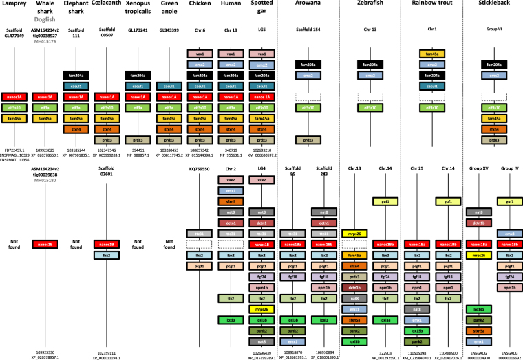
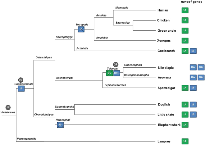
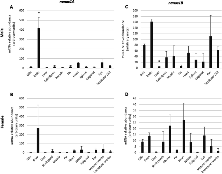
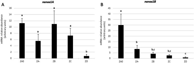
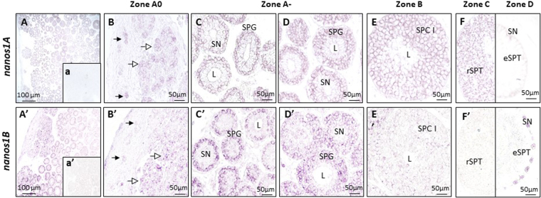
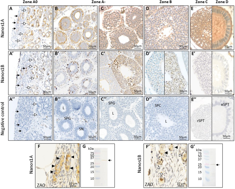
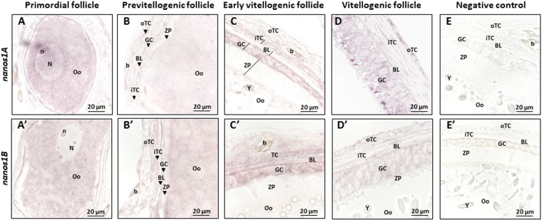
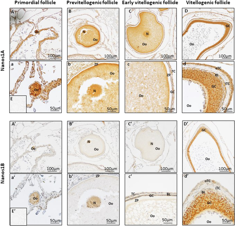
Nanos are RNA-binding proteins playing crucial roles in germ cell development and maintenance. Based on phylogenetic and synteny analyses, this study reveals that nanos1 gene has undergone multiple duplications and gene copies losses in Vertebrates. Chondrichthyan species display two nanos1 genes (named nanos1A/1B), which were both retrieved in some Osteichthyes at basal positions in Sarcopterygii and Actinopterygii lineages. In contrast, Teleosts have lost nanos1A but duplicated nanos1B leading to the emergence of two ohnologs (nanos1Ba/1Bb), whereas Tetrapods have lost nanos1B gene. The two successive nanos gene duplications may result from the second and third whole genome duplication events at the basis of Vertebrates and Teleosts respectively. The expression profiles of nanos1A and nanos1B paralogs were characterized in the dogfish, Scyliorhinus canicula. Nanos1A was strongly expressed in brain and also localized in all germ cell types in the polarized testis. In contrast, nanos1B was detected in testis with the highest expression in the germinative zone. In addition, Nanos1B protein was predominantly located in the nuclei of male germinal cells. In the ovary, both paralogs were detected in germinal and somatic cells. Our study opens new perspectives concerning the complex evolution of nanos1 paralogs and their potential distinct roles in Vertebrates gonads.


Conflict of interest statement

The authors declare no competing interests.

Figures

Figure 1
Figure 1
Gene synteny comparisons provide evidences that multiple nanos1 gene duplications and losses occurred in vertebrates Three nanos1 paralogs and their neighbouring genes showed syntenic genomic locations through Vertebrates. In the top panel, genes in the vicinity of a first copy, named nanos1A, were mapped. The figure was not drawn to scale. Each gene was represented by a specific coloured box. The name of each scaffold or chromosome harbouring the synteny is indicated at the top for each species whereas nanos1 gene copy and protein accession numbers are detailed at the bottom. Although the structure of the chromosomal fragment was well conserved during the evolution, the nanos1A gene copy is not observed in the genome of Teleost fish as symbolized by the dotted boxes. In the bottom panel, the genomic environment of the second nanos1 gene copy, termed nanos1B, was similarly represented. This different syntenic chromosomal fragment was not found in lamprey suggesting its apparition in Gnathostomata. Both nanos1 paralogs (nanos1A and nanos1B) were detected In Chondrichthyes (dogfish and whale shark) and in Osteichthyes, respectively at the basis of Sarcopterygii (coelacanth) and of Actinopterygii (spotted gar). In contrast, nanos1B gene was not found in elephant shark, xenopus, green anole, chicken and human, suggesting its loss in these species. Teleosts showed two nanos1B gene copies carried by similar but distinct chromosomal fragments. The two nanos1 paralogs in Teleosts were re-named nanos1Ba and nanos1Bb. Note that zebrafish is an atypical fish species because its genome does not harbour the nanos1Ba gene copy as indicated by a spotted box.
Figure 2
Figure 2
Hypothetical model of nanos1 gene duplications and copy losses during Vertebrate evolution. Based on nanos1 syntenies and phylogenetic trees, two rounds of duplication of the ancestral lamprey nanos1 gene were observed. At the basis of Vertebrates, the sea lamprey displays only the ancestral nanos1A gene copy. The first gene duplication giving rise to the nanos1B paralog may have occurred following the second genome duplication (2R) at the basis of Gnathostomata (box 1B). Chondrichthyan species (shark and ray) have kept both gene copies except the Holocephali (chimaera). In Osteichthyes, both paralogs were found in basal Sarcopterygii (coelacanth) but nanos1B was lost by Tetrapoda (crossed box 1B). Both nanos1A and nanos1B paralogs were found in basal Actinopterygii (spotted gar) but nanos1A was lost in Teleostei (crossed box 1A). Coinciding with the third round of whole genome duplication (3R), nanos1B gene copy would have undergone a second duplication in a Telelost ancestor giving rise to nanos1Ba and nanos1Bb gene copies (box 1Ba/b). The nanos1 paralogs present in the different species are summarized on the right.
Figure 3
Figure 3
nanos1A and nanos1B transcripts show distinct tissue distributions in male and female S. canicula. Messenger RNA relative abundances of nanos1A (A,B) and nanos1B (C,D) were quantified in panels of tissues in male (A,C) and female (B,D) dogfish by real-time PCR. Data were normalized using 5S rRNA. In both sexes, nanos1A showed a preferential expression in brain (A,B) whereas nanos1B showed a relatively ubiquitous expression pattern (C,D). Mature ovaries sampled from adult females and containing vitellogenic oocytes showed no significant difference of expression levels compared to immature ovaries sampled from pre-adult females and containing previtellogenic oocytes only (B,D). Data are presented as mean + SD (N = 3). Differences in gene expression were evaluated using one-way analysis of variance followed up with Games-Howell test. Significant differences (p ≤ 0.05) in gene expression compared to the testicular zone A0 or the immature ovary are indicated by asterisks (*).
Figure 4
Figure 4
Specific expression patterns of nanos1A and nanos1B in dogfish testicular zones. The relative abundance of nanos1A and nanos1B mRNAs in the five testicular zones was measured by RT-PCR and normalized with 5S rRNA. Dogfish polarized testis was dissected in five zones from its dorsal to its ventral side: ZA0, germinative zone containing SSCs; ZA-, cysts with spermatogonia; ZB, meiotic zone; ZC, cysts with round-spermatids; and ZD, cysts with elongated spermatids and zone of cyst resorption. The nanos1A transcript was detected in all zones except in zone D where it was almost undetectable. In contrast, nanos1B showed a marked progressive decrease of its expression after the germinative zone, ZA0. Data are presented as mean + SD (N = 4). Differences in gene expression were evaluated using one-way analysis of variance followed up with Games-Howell test. Testicular zones sharing no letter in common show significantly different expression levels.
Figure 5
Figure 5
Different but both germ cell-specific expressions of nanos1A and nanos1B in dogfish testis. The cellular distribution of nanos1 mRNAs was evaluated by in situ hybridization on dogfish testicular sections with antisense digoxigenin-conjugated riboprobes directed against nanos1A (AF) or nanos1B transcripts (A’–F’). a and a’ represent sense riboprobes ISH for nanos1A and nanos1B respectively. The nanos1A transcripts were detected in the potential SSC (filled arrow) and in undifferentiated spermatogonia (open arrow) of the zone A0 (B), in the spermatogonia (SPG) of zone A- (C,D) as well as in the primary spermatocytes (SPCI) of the zone B (E) and in the round spermatids (rSPT) of the zone C (F left panel). No signal was observed in the elongated spermatids (eSPT) of the zone D (F right panel) nor in the Sertoli cells (their nuclei are indicated by SN). The nanos1B transcripts were mainly detected in the potential SSC (filled arrow), the undifferentiated spermatogonia (open arrow) of zone A0 (B’) and in spermatogonia (SPG) of zone A- (C’,D’). A marked decrease in nanos1B expression was observed in the primary spermatocytes (SPCI) of zone B (E’) and no transcript was present in the germ cells of zone C (F’ left panel) and zone D (F’ right panel). Aspecific labelling was observed in Sertoli cells nuclei of zone D using nanos1B riboprobes (F’ right panel). L: lumen of the cyst; A, A’: overview of zone A of testis.
Figure 6
Figure 6
Different subcellular localizations of Nanos1A and Nanos1B proteins in dogfish testis. Immunohistochemistry has been performed on testis paraffin sections to detect Nanos1A (AF) and Nanos1B (A’–F’). 3,3′-diaminobenzidine (DAB) has been used for the revelation. A”–E” represent the negative control. The Nanos1A protein was detected in potential SSC (A filled arrow) and in undifferentiated spermatogonia (open arrow) leaving zone A0 (A). In these cells, Nanos1A localized in the cytoplasm (F filled arrowhead) and in the nucleus (F open arrowhead). The protein was also detectable during the different stages of spermatogenesis but only in germ cell cytoplasm as observed in zone A (B and C) in spermatogonia (SPG), in zone B (D) in spermatocytes (SPC), in zone C (E left panel) in round spermatids (rSPT) and finally in zone D (E right panel) in elongated spermatids (eSPT). In contrast, Nanos1B was expressed in potential SSCs (filled arrow, A’), undifferentiated spermatogonia (open arrow, A’) leaving the germinal niche and a fraction of differentiated spermatogonia (B’,C’) and spermatocytes (D’ right panel). No expression was detected in zones C (E left panel) and D (E right panel). Specific nucleus expression was noticed in some potential SSCs (F’ open arrowhead) and a specific DNA labelling was detected in spermatogonia and spermatocytes in division (A’–D’). The specificity of the antibodies used was validated by Western-blot analysis (G,G’). Male brain protein extract was used as nanos1 expression was the highest in this tissue. Proteins migrated in 15% SDS PAGE gels to separate low molecular weights since Nanos2 and Nanos3 theoretical molecular weights were estimated at 17.4 and 22.8 kDa respectively. Single bands were detected around the expected molecular weights, respectively 27.1 and 25 kDa for Nanos1A and Nanos1B.
Figure 7
Figure 7
Similar distribution of nanos1A and nanos1B transcripts in germ cells and in follicular cells in dogfish ovary. The localization of nanos1 mRNAs was determined in ovaries by in situ hybridization using antisense digoxigenin-conjugated riboprobes directed against nanos1A (A–D) or nanos1B transcripts (A’–D’). E and E’ represent results obtained with sense riboprobes for nanos1A and nanos1B respectively. The nanos1A transcripts were detected in primary oocyte of primordial follicles (around 100 µm in Ø illustrated in A). This labelling progressively decreased during oocyte growth until being undetectable in previtellogenic follicles (around 300 µm in Ø illustrated in (B). The mRNAs were also detected in the granulosa cells (GC) and the outer theca cells (oTC) of early (C) and the more advanced (D) vitellogenic follicles (around 500 µm and >2 mm in Ø respectively). The same expression profile was detected for nanos1B with an expression in primary oocyte of primordial follicles (A’), in granulosa (GC) and outer theca cells (oTC) of early (C) and more advanced (D) vitellogenic follicles. In previtellogenic follicle (B’), nanos1B was still detectable in oocyte cytoplasm. b: blood cells, BL: basal lamina, GC: granulosa cells, N: nucleus, n: nucleolus, Oo: primary oocyte, TC: theca cells, iTC: inner theca cells, oTC: outer theca cells, Y: yolk, ZP: zona pellucida.
Figure 8
Figure 8
Nanos1A and Nanos1B proteins share a similar expression profile in dogfish ovary. Immunohistochemistry has been performed on dogfish ovaries paraffin sections to detect Nanos1A (AD, higher magnification in a–d) and Nanos1B (A’–D’, higher magnification in a’–d’). 3,3′-diaminobenzidine (DAB) was used for the revelation. E and E’ illustrate the negative control. The Nanos1A protein was detected in primary oocytes from primordial follicles (around 100 µm in diameter, A,a) to early vitellogenic follicles (around 500 µm in Ø, C,c) in the cytoplasmic and nuclear (N) compartments. Staining in oocytes became progressively undetectable whereas it appeared in granulosa (GC) and outer theca cells (oTC) of early (C,c) and more advanced vitellogenic follicles (>2 mm in Ø, D,d). Similarly, Nanos1B was detected in oocytes of primordial follicles (A’,a’). In previtellogenic follicle (B’b’), Nanos1B presented a major expression in oocyte nucleus (N). Finally, Nanos1B was observed in granulosa cells (GC) and outer theca cells (oTC) in vitellogenic follicles (C’,c’ and D’,d’). BL: basal lamina, GC: granulosa cells, iTC: inner theca cells, N: nucleus, Oo: primary oocyte, oTC: outer theca cells, ZP: zona pellucida.

Similar articles

Cited by

References

    1. Arrizabalaga G, Lehmann R. A selective screen reveals discrete functional domains in Drosophila Nanos. Genetics. 1999;153:1825–1838. doi: 10.1093/genetics/153.4.1825. - DOI - PMC - PubMed
    1. Curtis D, et al. A CCHC metal-binding domain in Nanos is essential for translational regulation. EMBOI J. 1997;16:834–843. doi: 10.1093/emboj/16.4.834. - DOI - PMC - PubMed
    1. Hashimoto H, et al. Crystal structure of zinc-finger domain of Nanos and its functional implications. EMBO Rep. 2010;11:848–853. doi: 10.1038/embor.2010.155. - DOI - PMC - PubMed
    1. Suzuki A, Saga Y. Nanos2 suppresses meiosis and promotes male germ cell differentiation. Genes Dev. 2008;22:430–435. doi: 10.1101/gad.1612708. - DOI - PMC - PubMed
    1. Lehmann R, Nusslein-volhard C. The maternal gene nanos has a central role in posterior pattern formation of the Drosophila embryo. Development. 1991;112:679–691. doi: 10.1242/dev.112.3.679. - DOI - PubMed

Publication types

LinkOut - more resources