

Inherent flexibility of CLIC6 revealed by crystallographic and solution studies
- PMID: 29720717
- PMCID: PMC5931990
- DOI: 10.1038/s41598-018-25231-z
Inherent flexibility of CLIC6 revealed by crystallographic and solution studies
Abstract
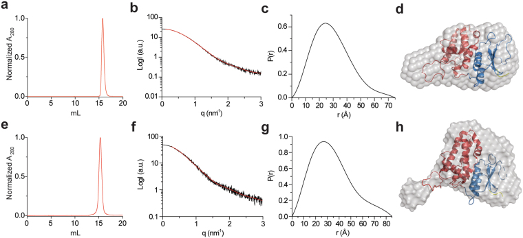
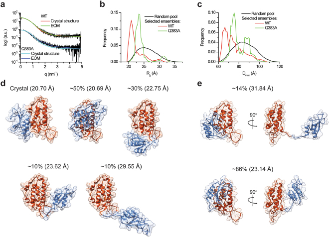
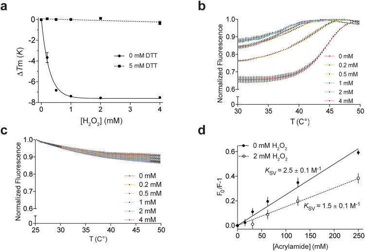
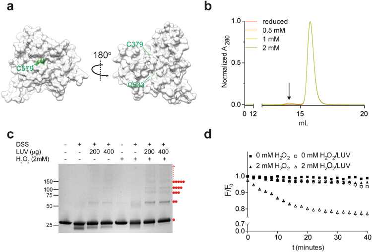
Chloride intracellular channels (CLICs) are a family of unique proteins, that were suggested to adopt both soluble and membrane-associated forms. Moreover, following this unusual metamorphic change, CLICs were shown to incorporate into membranes and mediate ion conduction in vitro, suggesting multimerization upon membrane insertion. Here, we present a 1.8 Å resolution crystal structure of the CLIC domain of mouse CLIC6 (mCLIC6). The structure reveals a monomeric arrangement and shows a high degree of structural conservation with other CLICs. Small-angle X-ray scattering (SAXS) analysis of mCLIC6 demonstrated that the overall solution structure is similar to the crystallographic conformation. Strikingly, further analysis of the SAXS data using ensemble optimization method unveiled additional elongated conformations, elucidating high structural plasticity as an inherent property of the protein. Moreover, structure-guided perturbation of the inter-domain interface by mutagenesis resulted in a population shift towards elongated conformations of mCLIC6. Additionally, we demonstrate that oxidative conditions induce an increase in mCLIC6 hydrophobicity along with mild oligomerization, which was enhanced by the presence of membrane mimetics. Together, these results provide mechanistic insights into the metamorphic nature of mCLIC6.
Conflict of interest statement
The authors declare no competing interests.
Figures

Similar articles
-
Structure and plasticity of the peptidyl-prolyl isomerase Par27 of Bordetella pertussis revealed by X-ray diffraction and small-angle X-ray scattering.J Struct Biol. 2010 Mar;169(3):253-65. doi: 10.1016/j.jsb.2009.11.007. Epub 2009 Nov 20. J Struct Biol. 2010. PMID: 19932182
-
Proteins at work: a combined small angle X-RAY scattering and theoretical determination of the multiple structures involved on the protein kinase functional landscape.J Biol Chem. 2010 Nov 12;285(46):36121-8. doi: 10.1074/jbc.M110.116947. Epub 2010 Aug 26. J Biol Chem. 2010. PMID: 20801888 Free PMC article.
-
The enigma of the CLIC proteins: Ion channels, redox proteins, enzymes, scaffolding proteins?FEBS Lett. 2010 May 17;584(10):2093-101. doi: 10.1016/j.febslet.2010.01.027. Epub 2010 Jan 19. FEBS Lett. 2010. PMID: 20085760 Review.
-
From glutathione transferase to pore in a CLIC.Eur Biophys J. 2002 Sep;31(5):356-64. doi: 10.1007/s00249-002-0219-1. Epub 2002 May 23. Eur Biophys J. 2002. PMID: 12202911 Review.
-
Hybrid Methods for Modeling Protein Structures Using Molecular Dynamics Simulations and Small-Angle X-Ray Scattering Data.Adv Exp Med Biol. 2018;1105:237-258. doi: 10.1007/978-981-13-2200-6_15. Adv Exp Med Biol. 2018. PMID: 30617833 Review.
Cited by
-
Chloride Intracellular Channel Proteins (CLICs) and Malignant Tumor Progression: A Focus on the Preventive Role of CLIC2 in Invasion and Metastasis.Cancers (Basel). 2022 Oct 6;14(19):4890. doi: 10.3390/cancers14194890. Cancers (Basel). 2022. PMID: 36230813 Free PMC article. Review.
-
Functional Relevance of Interleukin-1 Receptor Inter-domain Flexibility for Cytokine Binding and Signaling.Structure. 2019 Aug 6;27(8):1296-1307.e5. doi: 10.1016/j.str.2019.05.011. Epub 2019 Jun 27. Structure. 2019. PMID: 31257107 Free PMC article.
-
Inflammasomes as regulators of mechano-immunity.EMBO Rep. 2024 Jan;25(1):21-30. doi: 10.1038/s44319-023-00008-2. Epub 2023 Dec 15. EMBO Rep. 2024. PMID: 38177903 Free PMC article. Review.
-
The First Transcriptomic Atlas of the Adult Lacrimal Gland Reveals Epithelial Complexity and Identifies Novel Progenitor Cells in Mice.Cells. 2023 May 21;12(10):1435. doi: 10.3390/cells12101435. Cells. 2023. PMID: 37408269 Free PMC article.
-
A Literature-Derived Knowledge Graph Augments the Interpretation of Single Cell RNA-seq Datasets.Genes (Basel). 2021 Jun 10;12(6):898. doi: 10.3390/genes12060898. Genes (Basel). 2021. PMID: 34200671 Free PMC article.
References
-
- Argenzio, E. & Moolenaar, W. H. Emerging biological roles of Cl− intracellular channel proteins. J. Cell Sci. 129 (2016). - PubMed
Publication types
MeSH terms
Substances
LinkOut - more resources
Full Text Sources
Other Literature Sources
Molecular Biology Databases
