

Establishing Differences in Thermographic Patterns between the Various Complications in Diabetic Foot Disease
- PMID: 29721019
- PMCID: PMC5867599
- DOI: 10.1155/2018/9808295
Establishing Differences in Thermographic Patterns between the Various Complications in Diabetic Foot Disease
Abstract
Aim: To evaluate the potential of thermography as an assessment tool for the detection of foot complications by understanding the variations in temperature that occur in type 2 diabetes mellitus (DM).
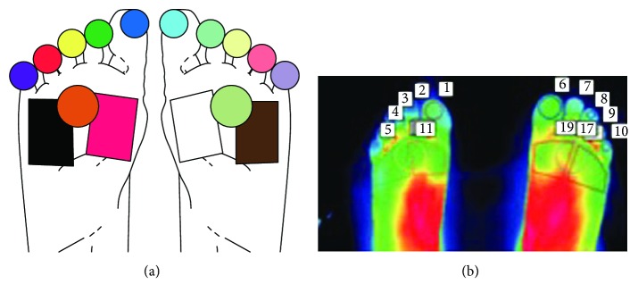
Methods: Participants were categorized according to a medical examination, ankle brachial index, doppler waveform analysis, and 10-gram monofilament testing into five groups: healthy adult, DM with no complications, DM with peripheral neuropathy, DM with neuroischaemia, and DM with peripheral arterial disease (PAD) groups. Thermographic imaging of the toes and forefeet was performed.
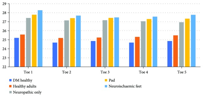
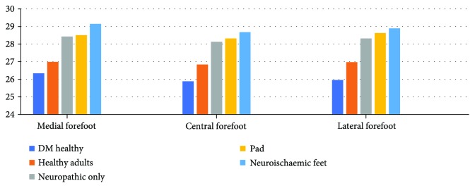
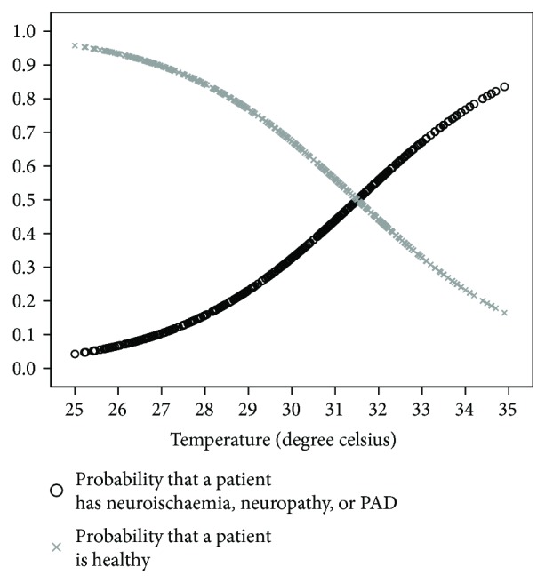
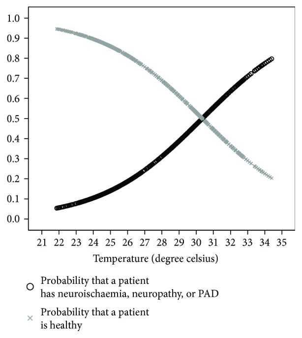
Results: 43 neuroischaemic feet, 41 neuropathic feet, 58 PAD feet, 21 DM feet without complications, and 126 healthy feet were analyzed. The temperatures of the feet and toes were significantly higher in the complications group when compared to the healthy adult and DM healthy groups. The higher the temperatures of the foot in DM, the higher the probability that it is affected by neuropathy, neuroischaemia, or PAD.
Conclusions: Significant differences in mean temperatures exist between participants who were healthy and those with DM with no known complications when compared to participants with neuroischaemia, neuropathy, or PAD. As foot temperature rises, so does the probability of the presence of complications of neuropathy, neuroischaemia, or peripheral arterial disease.
Figures
References
-
- Pinzur M. S., Slovenkai M. P., Trepman E., Shields N. N., Diabetes Committee of American Orthopaedic Foot and Ankle Society Guidelines for diabetic foot care: recommendations endorsed by the Diabetes Committee of the American Orthopaedic Foot and Ankle Society. Foot & Ankle International. 2005;26(1):113–119. doi: 10.1177/107110070502600112. - DOI - PubMed
-
- Bagavathiappan S., Philip J., Jayakumar T., et al. Correlation between plantar foot temperature and diabetic neuropathy: a case study by using an infrared thermal imaging technique. Journal of Diabetes Science and Technology. 2010;4(6):1386–1392. doi: 10.1177/193229681000400613. - DOI - PMC - PubMed
-
- Wang H., Wade D. R., Jr., Kam J. Burleigh D. D., Cramer K. E., Peacock G. R., editors. IR imaging of blood circulation of patients with vascular disease. Proc. SPIE. 2004. pp. 115–123. - DOI
LinkOut - more resources
Full Text Sources
Other Literature Sources
Research Materials
