. 2018 Mar 22;8(9):2348-2360.
doi: 10.7150/thno.22901. eCollection 2018.

A novel USP9X substrate TTK contributes to tumorigenesis in non-small-cell lung cancer

Affiliations

A novel USP9X substrate TTK contributes to tumorigenesis in non-small-cell lung cancer

Xiangling Chen et al. Theranostics. .

Abstract

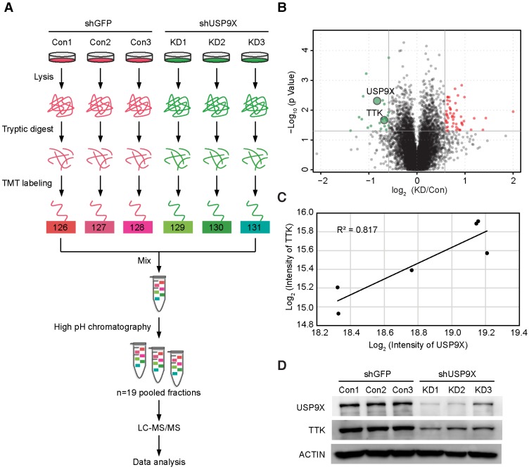
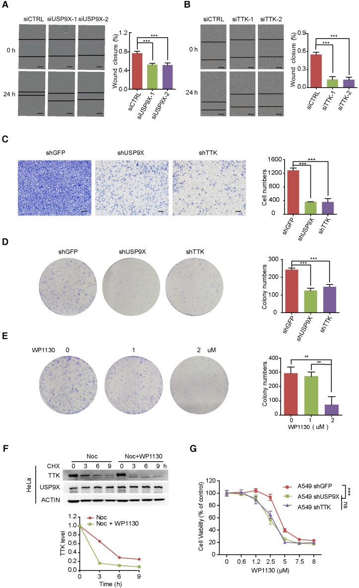
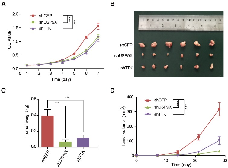
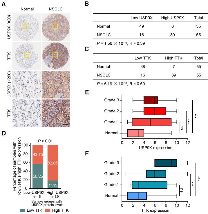
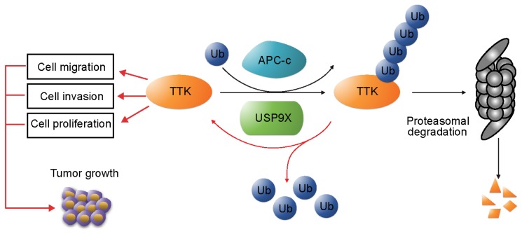
The X-linked deubiquitinase, USP9X, is implicated in multiple cancers by targeting various substrates. Increased expression of USP9X is observed in non-small-cell lung cancer (NSCLC) and is correlated with poor prognosis. However, the molecular mechanism for USP9X regulation of tumor cell survival and tumorigenesis in NSCLC is less defined. Methods: In this study, chemical labeling, quantitative proteomic screening was applied to analyze A549 cells with or without USP9X RNA interference. Functional in vitro and in vivo experiments were performed to confirm the oncogenic effects of USP9X in NSCLC and to investigate the underlying mechanisms. Results: The resulting data suggested that dual specificity protein kinase TTK is a potential substrate of USP9X. Further experimental evidences confirmed that USP9X stabilized TTK via direct interaction and efficient deubiquitination of TTK on K48 ubiquitin chain. Moreover, knockdown of USP9X or TTK inhibited cell proliferation, migration and tumorigenesis, and the immunohistochemical analysis of clinical NSCLC samples showed that the protein expression levels of USP9X and TTK were significantly elevated and positively correlated in tumor tissues. Conclusions: In summary, our data demonstrated that the USP9X-TTK axis may play a critical role in NSCLC, and could be considered as a potential therapeutic target.

Keywords: TTK; USP9X; deubiquitinase; non-small cell lung cancer (NSCLC); quantitative proteomics.


Conflict of interest statement

Competing Interests: The authors have declared that no competing interest exists.

Figures

Figure 1
Figure 1
TMT-based quantitative proteomics identifies TTK as a candidate substrate of USP9X. (A) Flow diagram of the TMT-based quantitative proteomics platform applied to identify the substrates of USP9X. A549 cells were stably transfected with 3 different shRNAs targeting USP9X (KD1, KD2, KD3) or control shRNA (Con1, Con2, Con3) and the whole cellular proteins were extracted and quantified. Following trypsin digestion of equal amount of proteins, the resolved peptides were labeled with 6-plex TMT reagents, fractionated by HPLC and analyzed by mass spectrometry. (B) Summary of the TMT labeling assay results. 7471 proteins identified by TMT assay are plotted in the volcano plot, in which the logarithmic ratio of protein intensities in the shUSP9X/control shRNA samples are plotted against negative logarithmic P values of the t-test performed from three replicates. 22 proteins were significantly down-regulated (green), 53 proteins were up-regulated (red) (fold change > 1.5, students' t test P value < 0.05). (C) Decreased USP9X expression correlates with decreased TTK protein level. (D)Validation of protein expression of USP9X and TTK in stably expressing A549 cells by immunoblotting.
Figure 2
Figure 2
USP9X is functionally linked to the stability of TTK. (A) GFP, empty vector control (EV), and flag-TTK were transfected into 293T cells. The cells were extracted for immunoprecipitation with the anti-flag agarose and the proteins were analyzed by immunoblotting. (B) Immunoprecipitated proteins with anti-USP9X antibodies or control IgG from 293T extracts were analyzed by western blotting. Endogenous TTK interacting with USP9X was detected using an anti-TTK antibody. (C) A549 cells were transfected with either two different siRNAs targeting USP9X or a control siRNA. After 72 h, cells were harvested and proteins were analyzed by immunoblotting. (D) The effect of USP9X depletion on TTK in HeLa cells as in (C). (E) Flag-TTK, HA-Ub, siRNA or siUSP9X were co-transfected into 293T cells. The cells were treated with or without MG132 as indicated. Then, cells were extracted for immunoprecipitation with anti-flag agarose and analyzed by western blotting. (F) Flag-TTK, ubiquitin K48-only plasmids, siRNA or siUSP9X were co-transfected into 293T cells. The indicated cells were treated with MG132 overnight. Then, cells were extracted for immunoprecipitation with anti-flag agarose and analyzed by western blotting. (G) A549 cells were transfected with either control or USP9X siRNAs. After 48 h, 200 μg/mL CHX was added and cells were harvested at the indicated times. Protein samples were analyzed by immunoblotting. Quantification of TTK levels relative to β-actin are presented. (H) Half-life analysis of TTK in constructed stable A549 cells. (I-J) HeLa (I) or 293FT (J) cells were transfected with plasmids expressing functional V5-USP9X (WT), catalytically dead C1566S (CS) USP9X or empty vector (EV). After 36 h, 200 μg/mL CHX was added and cells were harvested for immunoblotting.
Figure 3
Figure 3
Knock down of USP9X or TTK inhibits cell migration and invasion. (A) A549 cells were transfected with either two different siRNAs targeting USP9X or a control siRNA. After 48 h, cell migration was measured by a wound-healing assay. The wound edges are indicated by black lines. Representative images are shown. The quantitative results are shown on the right. The y-axis represents the percentage of wound closure. Data are represented as mean ± s.d. (n = 3, *** P < 0.001, t-test). Scale bars, 300 µM. (B) The migration ability of A549 cells with depletion of TTK examined via the wound-healing assay as in (A). Data are represented as mean ± s.d. (n = 3, *** P < 0.001, t-test). Scale bars, 300 µM. (C) Stable knockdown of either USP9X or TTK decreased A549 cells' invasion ability, which was measured by a transwell assay. The blue dye indicates the transwell cells. The quantitative results by counting cell numbers are shown on the right. Data are represented as mean ± s.d. (n = 3, *** P < 0.001, t-test). Scale bars, 100 µM. (D) Stable knockdown of either USP9X or TTK decreased A549 cells' colony forming ability, which was measured by a colony formation assay. The quantitative results by counting cell numbers are shown on the right. Data are represented as mean ± s.d. (n = 3, *** P < 0.001, t-test). (E) WP1130, an inhibitor of DUBs, could decrease A549 cells' colony forming ability. A549 cells were treated with the indicated drug concentration for the colony formation assay. After two weeks, cells were subjected to crystal violet staining. Data are represented as mean ± s.d. (n = 3, ** P < 0.01, t-test). (F) HeLa cells were induced into mitosis by treating with nocdazole, then the cells were treated with or without WP1130 as the indicated times and the cells were harvested for immunoblotting. Quantification of TTK levels relative to β-actin are shown. (G) Depletion of USP9X or TTK increased A549 cells' sensitivity to WP1130. A549 cells stably expressing shGFP, shUSP9X or shTTK were seeded into 96-well plates and then treated with different concentrations of WP1130 as indicated. The optical density (OD) at 490 nm for each well was detected. Data are represented as mean ± s.d. (n = 5, *** P < 0.001, t-test).
Figure 4
Figure 4
USP9X and TTK knockdown partially suppres cell proliferation and NSCLC. (A) A549 cells stably expressing shGFP, shUSP9X or shTTK were subjected to a cell proliferation assay. Each point represents the mean ± s.d. for biological quintuplicate experiments (n = 5, *** P < 0.001, two-way ANOVA). (B) Tumor images of nude mice (BALB/c; Slac Laboratory Animal, Shanghai, China) with subcutaneous injection of 5×10⁶ A549 cells stably transfected with shGFP, shUSP9X or shTTK at day 28 after implantation. (C) Weights of tumors in (B) were measured. Each bar represents the mean ± s.d. for different mice groups (n = 6, *** P < 0.001, one-way ANOVA). (D) Tumor volumes were measured weekly. Each bar represents the mean ± s.d. for different mice groups (n = 6, *** P < 0.001, two-way ANOVA).
Figure 5
Figure 5
The expression levels of USP9X and TTK are increased and correlated in NSCLC. (A) Representative immunohistochemical staining for USP9X in NSCLC tissues (n = 55) and matched adjacent normal tissues (n = 55). Scale bar, 50 μm. (B) USP9X expression levels in tumors and normal tissues. (C) TTK expression levels in tumors and normal tissues. Statistical significance in (B-C) was calculated by the chi-square test. R is the Pearson correlation coefficient. (D) Positive correlation between USP9X and TTK expression levels in NSCLC (** P < 0.01, chi-square test). (E) Correlation between USP9X protein expression level and the clinical stages of NSCLC. (F) Correlation between TTK protein expression level and the clinical stages of NSCLC (*** P< 0.001, one-way ANOVA).
Figure 6
Figure 6
USP9X-mediated stabilization of TTK promotes tumorigenesis in NSCLC. TTK is targeted for proteasome-dependent degradation by E3 ubiquitin ligase APC-c. USP9X decreases ubiquitination of TTK, and thereby enhances its half-life. Overexpression of USP9X would increase TTK levels and subsequently promote cell proliferation, migration and invasion. Immunohistochemistry experiments on clinical samples of NSCLC showed that elevated USP9X in NSCLC patients dramatically increases TTK levels and thus promotes tumor growth.

References

    1. Global Burden of Disease Cancer C, Fitzmaurice C, Dicker D, Pain A, Hamavid H, Moradi-Lakeh M. et al. The Global Burden of Cancer 2013. JAMA Oncol. 2015;1:505–27. - PMC - PubMed
    1. Ridge CA, McErlean AM, Ginsberg MS. Epidemiology of lung cancer. Semin Intervent Radiol. 2013;30:93–8. - PMC - PubMed
    1. Herbst RS, Heymach JV, Lippman SM. Lung cancer. N Engl J Med. 2008;359:1367–80. - PMC - PubMed
    1. Komander D, Rape M. The ubiquitin code. Annu Rev Biochem. 2012;81:203–29. - PubMed
    1. Komander D, Clague MJ, Urbe S. Breaking the chains: structure and function of the deubiquitinases. Nat Rev Mol Cell Biol. 2009;10:550–63. - PubMed

Publication types

MeSH terms