. 2018 Apr 10;9(27):19079-19099.
doi: 10.18632/oncotarget.24901.

An epigenetic regulator-related score (EpiScore) predicts survival in patients with diffuse large B cell lymphoma and identifies patients who may benefit from epigenetic therapy

Affiliations

An epigenetic regulator-related score (EpiScore) predicts survival in patients with diffuse large B cell lymphoma and identifies patients who may benefit from epigenetic therapy

Vanessa Szablewski et al. Oncotarget. .

Abstract

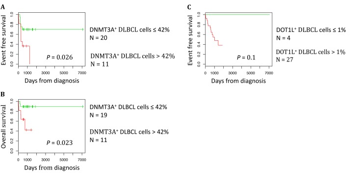
Diffuse large B-cell lymphoma (DLBCL) is the most common form of lymphoma and shows considerable clinical and biological heterogeneity. Much research is currently focused on the identification of prognostic markers for more specific patients' risk stratification and on the development of therapeutic approaches to improve the long-term outcome. Epigenetic alterations are involved in various cancers, including lymphoma. Interestingly, epigenetic alterations are reversible and drugs to target some of them have been developed. In this study, we demonstrated that the gene expression profile of epigenetic regulators has a prognostic value in DLBCL and identified pathways that could be involved in DLBCL poor outcome. We then designed a new risk score (EpiScore) based on the gene expression level of the epigenetic regulators DNMT3A, DOT1L, SETD8. EpiScore was predictive of overall survival in DLBCL and allowed splitting patients with DLBCL from two independent cohorts (n = 414 and n = 69) in three groups (high, intermediate and low risk). EpiScore was an independent predictor of survival when compared with previously described prognostic factors, such as the International Prognostic Index (IPI), germinal center B cell and activated B cell molecular subgroups, gene expression-based risk score (GERS) and DNA repair score. Immunohistochemistry analysis of DNMT3A in 31 DLBCL samples showed that DNMT3A overexpression (>42% of positive tumor cells) correlated with reduced overall and event-free survival. Finally, an HDAC gene signature was significantly enriched in the DLBCL samples included in the EpiScore high-risk group. We conclude that EpiScore identifies high-risk patients with DLBCL who could benefit from epigenetic therapy.

Keywords: Diffuse large B cells lymphoma; epigenetics; gene expression profiling; prognostic value; targeted treatment.


Conflict of interest statement

CONFLICTS OF INTEREST The authors declare no conflicts of interest.

Figures

Figure 1
Figure 1. Epigenetic regulators with prognostic value in patients with DLBCL (Lenz R-CHOP cohort n = 233)
For a given gene, a prognostic expression cut-off was calculated using the Maxstat algorithm, as described in Materials and Methods, to split patients in two groups (high and low risk) according to their overall survival (OS). BRD1: bromodomain containing 1; CARM1: coactivator associated arginine methyltransferase 1; BRPF3: bromodomain and PHD finger containing 3; CDYL: chromodomain Y like; DNMT3A: DNA cytosine-5-methyltransferase 3 alpha; DOT1L: DOT1-like histone H3K79 methyltransferase; HDAC2: histone deacetylase 2; PRMT5: protein arginine methyltransferase 5; SETD8: also known as KMT5 lysine (K)-specific methyltransferase 5A; SP140: SP140 nuclear body protein.
Figure 2
Figure 2. Epigenetic gene expression in the ABC and GC DLBCL subgroups (Lenz R-CHOP and CHOP cohorts)
CDYL: chromodomain Y-like; DNMT3A: DNA cytosine-5-methyltransferase 3 alpha; DOT1L: DOT1-like histone H3K79 methyltransferase; PRMT5: protein arginine methyltransferase 5; SP140: SP140 nuclear body protein; ABC: activating B cell; GC: germinal center. Results were compared using Student t test. The box-plot diagrams included the median value and the interquartile rage (IQR). The error bars represent the minimum for the values under the median and the outliers are identified as the third quartile plus 1.5 IQR (SPSS software).
Figure 3
Figure 3. DNMT3A, SETD8 and DOT1L gene expression in DLBCL samples compared with normal centrocytes and centroblasts (GSE56315 dataset)
Results were compared using Student t test. The box-plot diagrams included the median value and the interquartile rage (IQR). The error bars represent the minimum for the values under the median and the outliers are identified as the third quartile plus 1.5 IQR (SPSS software).
Figure 4
Figure 4
Immunohistochemical analysis of DNMT3A expression in tonsillar lymphoid tissue (A and B) and in two patients with DLBCL at diagnosis (C and D) and immunohistochemical analysis of DOT1L expression in tonsillar lymphoid tissue (E and F) and in two patients with DLBCL at diagnosis (G and H). (A) Nuclear expression of DNMT3A in reactive T cells of the interfollicular region (blue arrow) in a reactive human tonsil sample. (B) Nuclear expression of DNMT3A in some naive B cells in the mantle zone (green arrow), and in tingible-body macrophages and follicular dendritic cells in the germinal center (red arrow) in a reactive human tonsil sample. (C) Immunohistochemical analysis of DNMT3A expression in a patient with DLBCL (IPI 3). The patient was in complete remission after treatment with R-CHOP (six cycles) without relapse after 2 years of follow-up. No expression in large tumoral B cells (blue arrow and inset) and nuclear expression in reactive T cells show (red arrow). (D) Strong nuclear expression of DNMT3A in malignant B cells (see also inset) in a patient with DLBCL (IPI 3) with disease progression despite R-CHOP treatment. The patient died of the disease 6 months the after initial diagnosis. (E–F) Nuclear expression of DOT1L in some centrocytes and centroblasts in the germinal center (red arrow) in a reactive human tonsil sample whereas naive B cells in the mantle zone (green arrow) are negative. (G) Strong nuclear expression of DOT1L in a patient with DLBCL (80% of tumor cells are positive). (H) Low nuclear expression of DOT1L in another patient with DLBCL (only 2% of tumor cells are positive).
Figure 5
Figure 5. DNMT3A protein expression is a prognostic factor in DLBCL
The prognostic information provided by DNMT3A protein expression was investigated using the Maxstat algorithm. A cut-off of 42% of stained tumor cells allowed splitting the patients in a high risk and low risk group (A) Event free survival, (B) Overall survival. The prognostic information provided by DOT1L protein expression was investigated using the Maxstat algorithm. A cut-off of 1% of stained tumor cells allowed splitting the patients in EFS high risk and low risk groups (C).
Figure 6
Figure 6. EpiScore predicts the overall survival in patients with DLBCL
(A) Kaplan-Meier estimates of the overall survival in patients from the Lenz R-CHOP cohort subdivided in four groups on the basis of DNMT3A, DOT1L and SETD8 gene expression. When two consecutive groups showed no significant difference, they were merged. (B) This process led to the identification of three groups: low risk (low expression of DNMT3A, DOT1L and SETD8; blue), intermediate risk (high expression of one of these three genes; green) and high risk (high expression of two or all three genes; red). (C–D) EpiScore prognostic value at diagnosis was confirmed in two other independent cohorts.
Figure 7
Figure 7. HDACi treatment induces toxicity in cell lines overexpressing EpiScore genes
DLBCL cell lines overexpressing 2 out of the 3 genes of the EpiScore (RI1 and NUDUL1) and SUDHL5, characterized by low expression of the 3 genes, were cultured for 10 days without drug (control), with 0.5 µM or with 1 µM of SAHA and cell viability was analyzed by trypan blue assay. Data are representative of three idependent experiments. Statistical significance was tested using a Wilcoxon test for pairs (*P < 0.05).

Similar articles

Cited by

References

    1. Swerdlow S, Campo E, Harris N, Jaffe E, Pileri S, Stein H, Thiele J, Vardiman J. WHO Classification of Tumours of Haematopoietic and Lymphoid Tissues, Fourth Edition
    1. Blay J, Gomez F, Sebban C, Bachelot T, Biron P, Guglielmi C, Hagenbeek A, Somers R, Chauvin F, Philip T. The International Prognostic Index correlates to survival in patients with aggressive lymphoma in relapse: analysis of the PARMA trial. Parma Group. Blood. 1998;92:3562–8. - PubMed
    1. Alizadeh AA, Eisen MB, Davis RE, Ma C, Lossos IS, Rosenwald A, Boldrick JC, Sabet H, Tran T, Yu X, Powell JI, Yang L, Marti GE, et al. Distinct types of diffuse large B-cell lymphoma identified by gene expression profiling. Nature. 2000;403:503–11. https://doi.org/10.1038/35000501. - DOI - PubMed
    1. Hans CP, Weisenburger DD, Greiner TC, Gascoyne RD, Delabie J, Ott G, Müller-Hermelink HK, Campo E, Braziel RM, Jaffe ES, Pan Z, Farinha P, Smith LM, et al. Confirmation of the molecular classification of diffuse large B-cell lymphoma by immunohistochemistry using a tissue microarray. Blood. 2004;103:275–82. https://doi.org/10.1182/blood-2003-05-1545. - DOI - PubMed
    1. Sjö LD, Poulsen CB, Hansen M, Møller MB, Ralfkiaer E. Profiling of diffuse large B-cell lymphoma by immunohistochemistry: identification of prognostic subgroups. Eur J Haematol. 2007;79:501–7. https://doi.org/10.1111/j.1600-0609.2007.00976.x. - DOI - PubMed