Review
. 2018 Feb 23;5(1):3-25.
doi: 10.1002/reg2.97. eCollection 2018 Mar.

The diversity of myeloid immune cells shaping wound repair and fibrosis in the lung

Affiliations
Review

The diversity of myeloid immune cells shaping wound repair and fibrosis in the lung

Laura Florez-Sampedro et al. Regeneration (Oxf). .

Abstract

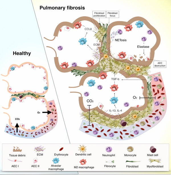
In healthy circumstances the immune system coordinates tissue repair responses in a tight balance that entails efficient inflammation for removal of potential threats, proper wound closure, and regeneration to regain tissue function. Pathological conditions, continuous exposure to noxious agents, and even ageing can dysregulate immune responses after injury. This dysregulation can lead to a chronic repair mechanism known as fibrosis. Alterations in wound healing can occur in many organs, but our focus lies with the lung as it requires highly regulated immune and repair responses with its continuous exposure to airborne threats. Dysregulated repair responses can lead to pulmonary fibrosis but the exact reason for its development is often not known. Here, we review the diversity of innate immune cells of myeloid origin that are involved in tissue repair and we illustrate how these cell types can contribute to the development of pulmonary fibrosis. Moreover, we briefly discuss the effect of age on innate immune responses and therefore on wound healing and we conclude with the implications of current knowledge on the avenues for future research.

Keywords: dendritic cells; fibrocytes; macrophages; monocytes; neutrophils.


Figures

Figure 1
Figure 1
Phases of tissue repair in the context of myeloid cells. (1) Clotting (top right) is the first step after injury takes place. Damage to alveolar epithelial cells (AEC) leads to the aggregation of erythrocytes and platelets that form a blood clot to contain spreading of the damage. The destruction of the tissue causes release of damage‐associated molecular patterns (DAMPs), or allows the entrance of microorganisms and thereby pathogen‐associated molecular patterns (PAMPs). Local cells such as dendritic cells, macrophages, and AECs are activated by DAMPs and PAMPs through toll‐like receptors (TLRs) and protease‐activated receptors (PARs) and produce the first round of pro‐inflammatory mediators. (2) Inflammation (bottom right) involves the infiltration of neutrophils and monocytes that respond to the pro‐inflammatory stimuli that were produced by resident lung cells. Phagocytes such as neutrophils and macrophages remove tissue debris and potentially threatening particles. In this pro‐inflammatory environment, monocytes and resident macrophages can differentiate into M1 macrophages that further promote inflammatory responses by the production of pro‐inflammatory mediators such as IL‐6 and TNF‐α. (3) Repair (bottom left) is the phase in which inflammatory responses subside and turn into repair responses through the effects of anti‐inflammatory and pro‐repair cytokines such as IL‐10 and TGF‐β. In this stage fibrocytes enter the tissue and differentiate into fibroblasts that proliferate and turn into the more contractile myofibroblasts. Myofibroblasts produce extracellular matrix (ECM) to close the open wound and form a scar. M2 macrophages predominate in this stage and produce mediators that contribute to the proliferation of fibroblasts and the deposition of ECM. (4) Resolution (top left) refers to the last phase in tissue repair in which excess ECM is degraded to make space for new cells. At the end of this stage the tissue has regained its structure and function
Figure 2
Figure 2
Pathophysiology of pulmonary fibrosis in the context of myeloid cells. The pathogenesis of pulmonary fibrosis is characterized by an exaggerated repair response to lung injury. These alterations in repair responses include proliferation of fibroblasts in the area of the injury and differentiation towards myofibroblasts that can form fibroblast foci. Dendritic cells accumulate in fibroblast foci in an immature form due to the influence of fibroblasts, which decreases T cell activation and proliferation. The predominant Th2 cytokine profile (e.g., IL‐4, IL‐13), produced by mast cells among others, promotes the polarization of macrophages towards an M2 phenotype. In turn, M2 macrophages produce soluble mediators such as TGF‐β and CCL8 that lead to fibroblast proliferation and to their differentiation into myofibroblasts and subsequent production of extracellular matrix (ECM). Moreover, deposition of ECM stimulates CCL8 production by alveolar macrophages resulting in a profibrotic vicious cycle. High numbers of neutrophils in fibrotic lung tissue contribute to the fibrotic process by perpetuating tissue damage and epithelial destruction via the production of elastase and possibly also by NETosis. High percentages of circulating fibrocytes are characteristic of pulmonary fibrosis. These circulating fibrocytes enter lung tissue and develop into fibroblasts that in the fibrotic environment differentiate into myofibroblasts. Additionally, fibrocytes contribute to the fibrotic process by producing TGF‐β and other mediators that contribute to the redundant and uncontrolled pro‐repair environment of the fibrotic lung

References

    1. Acharya, K. R. , & Ackerman, S. J. (2014). Eosinophil granule proteins: form and function. Journal of Biological Chemistry, 289(25), 17406–17415. - PMC - PubMed
    1. Adhyatmika, A. , Putri, K. S. S. , Beljaars, L. , & Melgert, B. N. (2015). The elusive antifibrotic macrophage. Frontiers in Medicine, 2(11), 81. - PMC - PubMed
    1. Aldenborg, F. , Nilsson, K. , Jarlshammar, B. , Bjermer, L. , & Enerback, L. (1993). Mast cells and biogenic amines in radiation‐induced pulmonary fibrosis. American Journal of Respiratory Cell and Molecular Biology, 8(1), 112–117. - PubMed
    1. Andersson, C. K. , Mori, M. , Bjermer, L. , Lofdahl, C. G. , & Erjefalt, J. S. (2010). Alterations in lung mast cell populations in patients with chronic obstructive pulmonary disease. American Journal of Respiratory and Critical Care Medicine, 181(3), 206–217. - PubMed
    1. Andersson‐Sjöland, A. , Garc, C. , Nihlberg, K. , Becerril, C. , Ram, R. , Pardo, A. , … Selman, M. (2008). Fibrocytes are a potential source of lung fibroblasts in idiopathic pulmonary fibrosis. International Journal of Biochemistry and Cell Biology, 40(10), 2129–2140. - PubMed

LinkOut - more resources