. 2018 Feb 1;7(5):e1424677.
doi: 10.1080/2162402X.2018.1424677. eCollection 2018.

CD96 targeted antibodies need not block CD96-CD155 interactions to promote NK cell anti-metastatic activity

Affiliations

CD96 targeted antibodies need not block CD96-CD155 interactions to promote NK cell anti-metastatic activity

Amelia Roman Aguilera et al. Oncoimmunology. .

Abstract

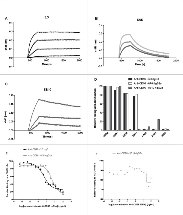
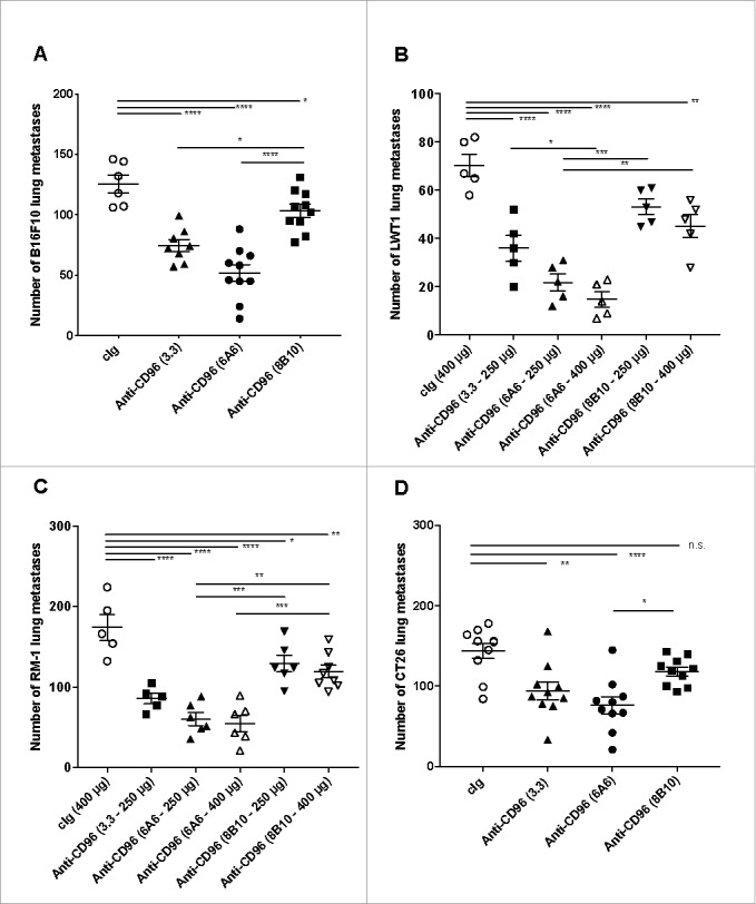
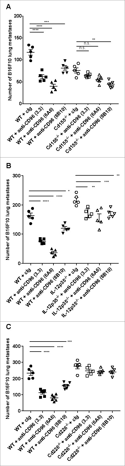
CD96 is a transmembrane glycoprotein Ig superfamily receptor, expressed on various T cell subsets and NK cells, that interacts with nectin and nectin-like proteins, including CD155/polio virus receptor (PVR). Here, we have compared three rat anti-mouse CD96 mAbs, including two that block CD96-CD155 (3.3 and 6A6) and one that does not block CD96-CD155 (8B10). Using flow cytometry, we demonstrated that both mAbs 3.3 and 6A6 bind to the first Ig domain of mouse CD96 and compete with CD155 binding, while mAb 8B10 binds to the second Ig domain and does not block CD155. While Fc isotype was irrelevant concerning the anti-metastatic activity of 3.3 mAb, in four different experimental metastases models and one spontaneous metastasis model, the relative order of anti-metastatic potency was 6A6 > 3.3 > 8B10. The metastatic burden control of all of the anti-CD96 clones was highly dependent on NK cells and IFN-γ. Consistent with its inability to block CD96-CD155 interactions, 8B10 retained anti-metastatic activity in CD155-deficient mice, whereas 3.3 and 6A6 lost potency in CD155-deficient mice. Furthermore, 8B10 retained most of its anti-metastatic activity in IL-12p35-deficient mice whereas the activity of 3.3 and 6A6 were partially lost. All three mAbs were inactive in CD226-deficient mice. Altogether, these data demonstrate anti-CD96 need not block CD96-CD155 interactions (ie. immune checkpoint blockade) to promote NK cell anti-metastatic activity.

Keywords: CD155; CD96; NK cells; immunotherapy; metastasis.


Figures

Figure 1.
Figure 1.
Anti- mCD96 mAbs bind different Ig domains of the CD96 molecule. Octet binding profiles of (A) 3.3, (B) 6A6 and (C) 8B10 anti-CD96 mAbs to recombinant CD96. (D) Relative binding of anti-mCD96 mAbs (3.3, 6A6 and 8B10) to mouse/human CD96 three domain chimeric molecules (M = mouse and H = human) expressed in HEK-293 cells as measured by flow cytometry where binding to mouse CD96 is standardized as 100%. Relative binding curves of mCD155-Fc (3 μg/ml) binding to HEK-293 cells transfected with mCD96 in the presence of various dilutions (log concentration shown starting at 100 μg/ml) (E) 3.3 and 6A6 mAbs and (F) 8B10 mAb as indicated.
Figure 2.
Figure 2.
Dose response of anti-CD96 mAbs, against B16F10 experimental lung metastases. WT mice were injected i.v. with B16F10 melanoma (1 × 10⁵ cells). On day 0 and 3 after tumor inoculation, mice were treated with i.p. injections of cIg (250 μg) or various doses of anti-CD96 mAb (A) 6A6 (rat IgG2a) and (B) 8B10 (rat IgG2a) as indicated. The cIg group shown is common to parts A and B. The metastatic burden was quantified in the lungs after 14 days by counting colonies on the lung surface. Individual mice are represented by each symbol and means ± SEM of 5 mice per group are shown. Significant differences between groups as indicated by crossbars were determined by a one-way ANOVA with post Tukey test (*: p < 0.05, **:p < 0.01, ****: p < 0.0001).
Figure 3.
Figure 3.
Anti-CD96 mAbs differentially suppress experimental lung metastasis. Comparative activities of various anti-CD96 mAbs against various experimental lung metastases. Groups of 5–10 C57BL/6 or BALB/c WT were injected i.v. with (A) B16F10 melanoma (1 × 10⁵cells), (B) LWT1 melanoma (5 × 10⁵ cells), (C) RM-1 prostate carcinoma (5 × 10⁴ cells), or (D) CT26 colon adenocarcinoma (5 × 10⁵ cells). On days 0 and 3 after tumor inoculation, mice were treated with i.p. injections of cIg (400 μg), anti-CD96 mAb 3.3 (250 μg, rat IgG1), 6A6 (250 or 400 μg, rat IgG2a), or 8B10 (400 μg, rat IgG2a), as indicated. The metastatic burden was quantified in the lungs after 14 days by counting colonies on the lung surface. Individual mice are represented by each symbol and means ± SEM of 5–10 mice per group are shown. Significant differences between groups as indicated by crossbars were determined by a one-way ANOVA with post Tukey test (*: p < 0.05, **: p < 0.01, ***: p < 0.001; ****: p < 0.0001). Part of panel A has already been published in Cancer Discovery article.
Figure 4.
Figure 4.
Anti-CD96 mAbs differentially suppress spontaneous metastasis. Groups of 5–10 BALB/c WT mice were injected with 5 × 10⁴ 4T1.2 mammary carcinoma cells into the mammary fat pad. Some groups of mice were treated i.p. on days 12 and 14 after tumor inoculation with neoadjuvant cIg (400 μg), anti-CD96 mAb 3.3 (400 μg, rat IgG1), 6A6 (400 μg, rat IgG2a), 8B10 (400 μg, rat IgG2a), or anti-PD-1 (RMP1-14, 250 μg) as indicated. On days 16 all primary tumors were resected. (A) Some cohorts were sacrificed on day 34 and the metastatic burden was quantified in the lungs by counting colonies on the lung surface. Individual mice are represented by each symbol and means ± SEM of 5 mice per group are shown. Significant differences between groups as indicated by crossbars were determined by a one-way ANOVA with post Tukey test (*: p < 0.05, **: p < 0.01, ***: p < 0.001). (B) Other mice were monitored for survival. The Kaplan-Meier curves for overall survival of each group are shown. Significant differences between indicated groups were determined by log-rank sum test with exact p values shown.
Figure 5.
Figure 5.
Anti-CD96 mAb suppression of metastases is not dependent on Fc function. Groups of 4–8 C57BL/6 WT or various FcR-deficient mice (FcγRIII, FcγRIV or Fcεγ) were injected i.v. with B16F10 melanoma (1 × 10⁵cells). On days 0 and 3 after tumor inoculation, mice were treated with i.p. injections of (A-C) cIg (250 or 400 μg), (A) anti-CD96 mAb 3.3 (250 μg, rat IgG1), (B) 6A6 (250 μg, rat IgG2a), or (C) 8B10 (400 μg, rat IgG2a) as indicated. (D) Groups of 5 C57BL/6 WT mice were injected i.v. with B16F10 melanoma (2 × 10⁵cells). On days 0 and 3 after tumor inoculation, mice were treated with 250 μg i.p. injections of cIg (rat G1), 3.3 (rat IgG1), 3.3 (mouse IgG1), 3.3 (mouse IgG1 D265A), or 3.3 (mouse IgG2a). The metastatic burden was quantified in the lungs after 14 days by counting colonies on the lung surface. Individual mice are represented by each symbol and means ± SEM of 4–8 mice per group are shown. Significant differences between groups as indicated by crossbars were determined by a one-way ANOVA with post Tukey test (*: p < 0.05, **: p < 0.01, ***: p < 0.001).
Figure 6.
Figure 6.
Anti-CD96 suppression of experimental lung metastasis depends upon NK cells, CD96 and IFN-γ. C57BL/6 WT and CD96-deficient mice (Cd96−/−) mice as indicated were injected i.v with B16F10 melanoma (2 × 10⁵cells). On days 0 and 3 after tumor inoculation, mice were treated with i.p. injections of (A, B) cIg (250 μg) or anti-CD96 mAbs (A) 6A6 (250 μg) or (B) 8B10 (250 μg). Some groups of mice were treated i.p. on days −1, 0 and 7 after tumor inoculation with cIg (100 μg), anti-asGM1 (50 μg) or anti-IFN-γ (250 μg). The metastatic burden was quantified in the lungs after 14 days by counting colonies on the lung surface. Mean ± SEM of 5 mice per group are shown. A and B were the same experiment and some of the cIg and anti-asGM1 treated control groups are replicated in each panel for visual display. Significant differences between groups as indicated by crossbars were determined by a one-way ANOVA with post Tukey test (*: p < 0.05, **: p < 0.01, ****: p < 0.0001).
Figure 7.
Figure 7.
Anti-CD96 8B10 mAbs protects from metastases in the absence of CD155 and IL-12p35. (A) Groups of C57BL/6 WT or CD155-deficient (Cd155−/−) mice were injected i.v. with B16F10 melanoma (1 × 10⁵ cells). On days 0 and 3 after tumor inoculation, mice were treated with i.p. injections of anti-CD96 3.3 mAb (rat IgG1, 250 μg), 6A6 mAb (rat IgG2a, 250 μg), and 8B10 mAb (rat IgG2a, 250 μg) or the equivalent amount of cIg (250 μg). (B) Groups of C57BL/6 WT or IL-12p35-deficient (IL-12p35−/−) mice were injected i.v. with B16F10 melanoma (2 × 10⁵ cells). On days 0 and 3 after tumor inoculation, mice were treated with i.p. injections of anti-CD96 3.3 mAb (rat IgG1, 250 μg), 6A6 mAb (rat IgG2a, 250 μg), and 8B10 mAb (rat IgG2a, 250 μg) or the equivalent amount of cIg (250 μg). (C) Groups of C57BL/6 WT or Cd226-deficient (Cd226−/−) mice were injected i.v. with B16F10 melanoma (5 × 10⁵ cells). On days 0 and 3 after tumor inoculation, mice were treated with i.p. injections of anti-CD96 3.3 mAb (rat IgG1, 250 μg), 6A6 mAb (rat IgG2a, 250 μg), and 8B10 mAb (rat IgG2a, 250 μg) or the equivalent amount of cIg (250 μg). The metastatic burden was quantified in the lungs after 14 days by counting colonies on the lung surface. Mean ± SEM of 5–6 mice per group are shown. Significant differences between groups as indicated by crossbars were determined by a one-way ANOVA with post Tukey test (*: p < 0.05, **: p < 0.01, ***: p < 0.001; ****: p < 0.0001).

Similar articles

Cited by

References

    1. Christofori G. New signals from the invasive front. Nature. 2006;441:444–50. doi:10.1038/nature04872. - DOI - PubMed
    1. Gupta GP, Massague J. Cancer metastasis: building a framework. Cell. 2006;127:679–95. doi:10.1016/j.cell.2006.11.001. - DOI - PubMed
    1. Lopez-Soto A, Gonzalez S, Smyth MJ, Galluzzi L. Control of metastasis by NK cells. Cancer Cell. 2017;32:135–54. doi:10.1016/j.ccell.2017.06.009. - DOI - PubMed
    1. Blake SJ, Stannard K, Liu J, Allen S, Yong MC, Mittal D, Aguilera AR, Miles JJ, Lutzky VP, de Andrade LF, et al.. Suppression of metastases using a new lymphocyte checkpoint target for cancer immunotherapy. Cancer Discov. 2016;6:446–59. doi:10.1158/2159-8290.CD-15-0944. - DOI - PubMed
    1. Shibuya A, Campbell D, Hannum C, Yssel H, Franz-Bacon K, McClanahan T, Kitamura T, Nicholl J, Sutherland GR, Lanier LL, et al.. DNAM-1, a novel adhesion molecule involved in the cytolytic function of T lymphocytes. Immunity. 1996;4:573–81. doi:10.1016/S1074-7613(00)70060-4. - DOI - PubMed

Publication types