. 2018 Feb 8;7(5):e1426434.
doi: 10.1080/2162402X.2018.1426434. eCollection 2018.

Bufalin Suppresses Migration and Invasion of Hepatocellular Carcinoma Cells Elicited by Poly (I:C) Therapy

Affiliations

Bufalin Suppresses Migration and Invasion of Hepatocellular Carcinoma Cells Elicited by Poly (I:C) Therapy

Yinglu Feng et al. Oncoimmunology. .

Abstract

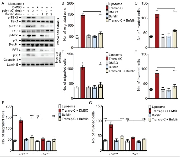
The Toll-like receptor 3 (TLR3) agonists as polyriboinosinic-polyribocytidylic acid (poly (I:C)) have been implicated as potential immunotherapy adjuvant for cancer whereas the exact roles of TLR3 agonists in hepatocellular carcinoma (HCC) treatment have not been clearly evaluated. In consistent with previous reports, we found that poly (I:C) triggering of TLR3 inhibited cell proliferation and induced apoptosis in HCC cells. However, poly (I:C), when used at lower concentration that cannot remarkably inhibit proliferation and induce apoptosis in HCC cells, enhanced the migration and invasion in vitro and the metastasis in vivo. More importantly, we found that bufalin, a prominent component of toad venom, could suppress poly (I:C)-inspired migration, invasion and metastasis of HCC cells despite that bufalin could not potentiate poly (I:C)-induced inhibition of proliferation and induction of apoptosis. In MHCC97 H cells, bufalin impaired poly (I:C)-induced activation of Tank-binding kinase 1 (TBK1) and interferon regulatory factor 3 (IRF3) pathway and NF-κB pathway. Inhibitor for TBK1 but not NF-κB suppressed poly (I:C)-inspired migration and invasion, which was further supported by using TBK1 deficient (Tbk1-/- ) cells. In another model using poly (I:C) transfection, bufalin could also suppress the migration and invasion of HCC cells, which was not observed in Tbk1-/- MHCC97 H cells. Our data suggest that bufalin can suppress the metastasis of HCC cells in poly (I:C) therapy by impairing TBK1 activation, indicating that bufalin may be used in combination with poly (I:C) therapy in HCC treatment for the sake of reversing poly (I:C)-triggered metastasis of HCC cells.

Keywords: Bufalin; HCC; TBK1; TLR3; metastasis; poly (I:C).


Figures

Figure 1.
Figure 1.
Effects of poly (I:C) and bufalin on cell proliferation and apoptosis of HCC cells. (A and B) Determination of TLR3 amounts in HCC cells by q-PCR (A) and Western blotting (B). HEK293 cells and human peripheral mononuclear cells (hPBMC) were used as negative and positive control, respectively. (C to E) MHCC97 H cells were treated with increasing doses of poly (I:C) (5, 10, 20 or 50 μg/ml), bufalin (5, 10, 20 or 50 nM) or vehicles as indicated for 48 h. Cell proliferation was determined by BrdU incorporation as evaluated using OD450 nm (C), and apoptosis was determined by annxin V/PI staining as evaluated by FACS (D and E). In (A), (C) and (E), data are means ± SD of triplicates. In (D), the proportion (%) numbers were shown. Data are representative of three independent experiments. ns, not significant (one-way ANOVA followed by Bonferroni multiple comparisons).
Figure 2.
Figure 2.
Effects of poly (I:C) and bufalin on migration and invasion of HCC cells. (A to D) MHCC97 H cells were treated with increasing doses of poly (I:C) (2, 5 or 10 μg/ml), bufalin (5, 10 or 20 nM) or vehicles as indicated for 48 H. Migrated cells (A and B) and invaded cells (C and D) were either photographed (A and C, 200 x magnification) or counted (cells of one filed out of 5 fields of membrane; B and D, 200 x magnification). (E to G) HepG2 cells stably transfected with TLR3-Myc vector (HepG2-TLR3) or empty vector (HepG2-Mock) were treated with poly (I:C) (5 μg/ml), bufalin (5 nM) or vehicles as indicated for 48 H, and were examined for migration and invasion as in (A to D). Expression of TLR3 was examined by Western blotting using anti-Myc antibody (E). In (B), (D), (F) and (G), data are means ± SD of triplicates. Data are representative of three independent experiments. ns, not significant; ***, P < 0.001 (one-way ANOVA followed by Bonferroni multiple comparisons).
Figure 3.
Figure 3.
Bufalin inhibits poly (I:C)-triggered activation of TBK1-IRF3 pathway in HCC cells. (A to E) MHCC97 H cells were treated with poly (I:C) (5 μg/ml), bufalin (5 nM) or vehicles (DMSO, 2hrs) as indicated. The amounts of indicated proteins contained in 100 μg whole cell extracts (WCL) were determined by ELISA assays. In (E), equal amounts of WCL or nuclear extracts were examined for indicated proteins by using Western blotting. (F and G) MHCC97 H cells were transiently transfected with Ifnb (F) or NF-κB (G) reporters for 24 H, and treated with poly (I:C) (5 μg/ml), bufalin (5 nM) or vehicles for 6 H. Reporter activation was measured by dual-luciferase activity assays. In (A to D), (F) and (G), data are means ± SD of triplicates. Data are representative of three independent experiments. ns, not significant; *, P < 0.05; **, P < 0.01; ***, P < 0.001 (one-way ANOVA followed by Bonferroni multiple comparisons).
Figure 4.
Figure 4.
Roles of TBK1 in poly (I:C)– and bufalin–regulated migration and invasion of HCC cells. (A to D) MHCC97 H (A and B) or HepG2-TLR3 (C and D) cells were pretreated with or without TPCA-1 (100 nM) or BX-795 (100 nM) for 30 min, and then treated with poly (I:C) (5 μg/ml), bufalin (5 nM) or vehicles as indicated for 48 H. Migrated cells (A and C) and invaded cells (B and D) were counted (cells of one filed out of 5 fields of membrane under 200 x magnification). (E to J) Tbk1+/+ or Tbk1–/– MHCC97 H cells (E to G) or Tbk1–/– MHCC97 H cells rescued by transfection with empty (Mock) or TBK1 expression vectors for 24 H (H to J) were treated with poly (I:C) (5 μg/ml), bufalin (5 nM) or vehicles as indicated for 48 H, and were examined for migration and invasion as in (A to D). Expression of TBK1 was examined by Western blotting using anti-TBK1 antibody (E and H). In (A to D), (F), (G), (I) and (J), data are means ± SD of triplicates. Data are representative of three independent experiments. ns, not significant; ***, P < 0.001 (one-way ANOVA followed by Bonferroni multiple comparisons).
Figure 5.
Figure 5.
Bufalin inhibits the activation of TBK1 and suppresses migration and invasion of HCC cells triggered by poly (I:C) transfection. (A to E) MHCC97 H (A to C) or HepG2 (D and E) cells were transfected with poly (I:C) (5 μg/ml), or treated with bufalin (5 nM) or vehicles as indicated for 1–2 H (A) or 48 H (B to E). The amounts of indicated proteins in equal amounts of whole cell extracts or nuclear extracts were examined by Western blotting (A). Migrated cells (B and D) and invaded cells (C and E) were counted (cells of one filed out of 5 fields of membrane under 200 x magnification). (F and G) Tbk1+/+ or Tbk1–/– MHCC97 H cells were were transfected with poly (I:C) (5 μg/ml), or treated with bufalin (5 nM) or vehicles as indicated for 48 H. Migration and invasion were determined as in (B to E). In (B to G), data are means ± SD of triplicates. Data are representative of three independent experiments. ns, not significant; ***, P < 0.001 (one-way ANOVA followed by Bonferroni multiple comparisons).
Figure 6.
Figure 6.
Bufalin inhibits poly (I:C)-inspired metastasis of HCC cells in vivo. (A and B) Balb/c nu/nu mice with MHCC97 H orthotopical xenografts were treated by intraperitoneal injection of indicated amounts of poly (I:C) (0.5, 1 or 2 mg/kg), bufalin (0.5, 1 or 2 mg/kg) or vehicles as indicated. For the combination treatments, bufalin was administrated at 0.5 mg/kg. On the third day after the second poly (I:C) treatment, liver were examined for apoptosis by TUNEL assays. The section stained with only the HRP-conjugated secondary antibody was used as negative control. The sections were photographed under 400 x magnification, and TUNEL positive cells under each field of 5 fields were counted. (C to F) After six weeks of treatment (0.5 mg/kg poly (I:C) or 0.5 mg/kg bufalin), the lungs were excised and examined for metastases by H&E staining. Typical metastases were shown in (C; 200 x magnification), and the metastases under each field of 5 fields were counted. The orthotopical xenografts examined were: MHCC97 H (C and D), HepG2-TLR3 (E) and Tbk1+/+ or Tbk1–/– MHCC97 H (F). In (B), (D), (E) and (F), data are means ± SD (n = 10 per group). In (A) and (C), bars indicate for 50 μm. Data are representative of three independent experiments. ns, not significant; ***, P < 0.001 (one-way ANOVA followed by Bonferroni multiple comparisons).
Figure 7.
Figure 7.
Bufalin inhibits poly (I:C)-induced phosphorylation of TBK1 in vivo. Balb/c nu/nu mice with MHCC97 H (A and B) or HepG2-TLR3 (C and D) orthotopical xenografts were treated by intraperitoneal injection of poly (I:C) (0.5 mg/kg), bufalin (0.5 mg/kg) or vehicles as indicated. On the third day after the second poly (I:C) treatment, liver were examined for phospho-TBK1 expression by immunohistochemistry (IHC) assays. The sections were photographed under 400 x magnification (A and C), and the staining was semiquantitatively scored. The section derived from poly (I:C) group and stained in the presence of blocking peptide was used as negative control. In (B) and (D), data are means ± SD (n = 10 per group). In (A) and (C), bars indicate for 50 μm. Data are representative of three independent experiments. ***, P < 0.001 (one-way ANOVA followed by Bonferroni multiple comparisons).

References

    1. Ferlay J, Soerjomataram I, Dikshit R, Eser S, Mathers C, Rebelo M, Parkin DM, Forman D, Bray F. Cancer incidence and mortality worldwide: sources, methods and major patterns in GLOBOCAN 2012. Int J Cancer. 2015;136:E359–86. doi:10.1002/ijc.29210. PMID: 25220842. - DOI - PubMed
    1. El-Serag HB. Hepatocellular carcinoma. N Engl J Med. 2011;365:1118–27. doi:10.1056/NEJMra1001683. PMID: 21992124. - DOI - PubMed
    1. Cao X. Self-regulation and cross-regulation of pattern-recognition receptor signalling in health and disease. Nat Rev Immunol. 2016;16:35–50. doi:10.1038/nri.2015.8. PMID: 26711677. - DOI - PubMed
    1. Galluzzi L, Vacchelli E, Eggermont A, Fridman WH, Galon J, Sautès-Fridman C, Tartour E, Zitvogel L, Kroemer G. Trial Watch: Experimental Toll-like receptor agonists for cancer therapy. Oncoimmunology. 2012;1:699–716. doi:10.4161/onci.20696. PMID: 22934262. - DOI - PMC - PubMed
    1. Talmadge JE, Adams J, Phillips H, Collins M, Lenz B, Schneider M, Chirigos M. Immunotherapeutic potential in murine tumor models of polyinosinic-polycytidylic acid and poly-L-lysine solubilized by carboxymethylcellulose. Cancer Res. 1985;45:1066–72. PMID: 3971361 - PubMed

Publication types