

The Retinal Circadian Clock and Photoreceptor Viability
- PMID: 29721962
- PMCID: PMC6003627
- DOI: 10.1007/978-3-319-75402-4_42
The Retinal Circadian Clock and Photoreceptor Viability
Abstract
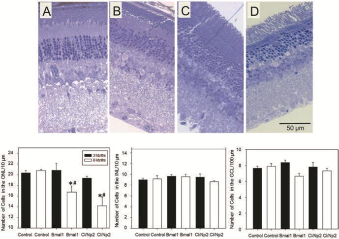
Circadian rhythms are present in most living organisms, and these rhythms are not just a consequence of the day/night fluctuation, but rather they are generated by endogenous biological clocks with a periodicity of about 24 h. In mammals, the master pacemaker of circadian rhythms is localized in the suprachiasmatic nuclei (SCN) of the hypothalamus. The SCN controls circadian rhythms in peripheral organs. The retina also contains circadian clocks which regulate many aspects of retinal physiology, independently of the SCN. Emerging experimental evidence indicates that the retinal circadian clocks also affect ocular health, and a few studies have now demonstrated that disruption of retinal clocks may contribute to the development of retinal diseases. Our study indicates that in mice lacking the clock gene Bmal1, photoreceptor viability during aging is significantly reduced. Bmal1 knockout mice at 8-9 months of age have 20-30% less nuclei in the outer nuclear layer. No differences were observed in the other retinal layers. Our study suggests that the retinal circadian clock is an important modulator of photoreceptor health.
Keywords: Aging; Cell viability; Circadian rhythm; Clock genes; Cone; Knockout mice; Oscillation; Photoreceptors; Retinal degeneration.
Figures
References
-
- Ait-Hmyed HO, Felder-Schmittbuhl MP, Garcia-Garrido M, et al. Mice lacking Period 1 and Period 2 circadian clock genes exhibit blue cone photoreceptor defects. Eur J Neurosci. 2013;37:1048–60. - PubMed
-
- Ait-Hmyed HO, Acar N, Savier E, et al. Rev-Erbα modulates retinal visual processing and behavioral responses to light. FASEB J. 2016 pii: fj.201600414R. - PubMed
Publication types
MeSH terms
Substances
Grants and funding
LinkOut - more resources
Full Text Sources
Other Literature Sources
Medical
