. 2018 Jul;175(14):2968-2987.
doi: 10.1111/bph.14346. Epub 2018 Jun 3.

Hippocampal PPARα is a novel therapeutic target for depression and mediates the antidepressant actions of fluoxetine in mice

Affiliations

Hippocampal PPARα is a novel therapeutic target for depression and mediates the antidepressant actions of fluoxetine in mice

Lu Song et al. Br J Pharmacol. 2018 Jul.

Abstract

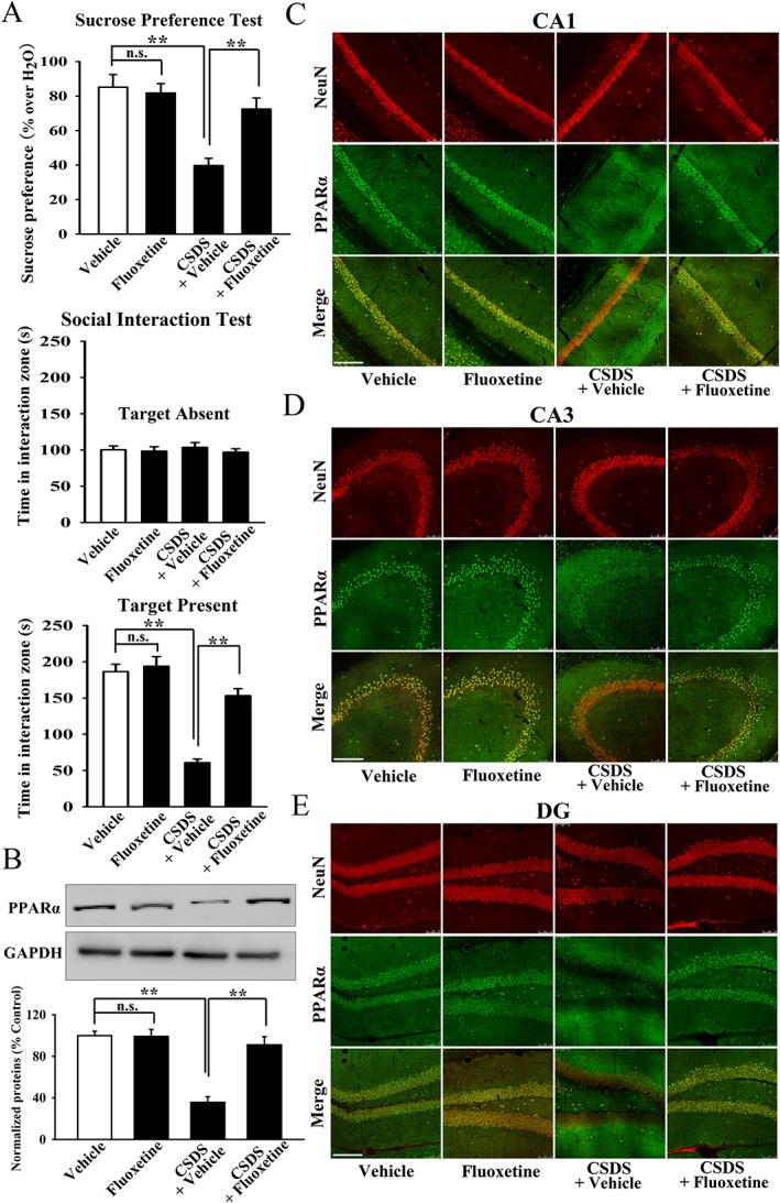
Background and purpose: Developing novel pharmacological targets beyond the monoaminergic system is now a popular strategy for treating depression. PPARα is a nuclear receptor protein that functions as a transcription factor,-regulating gene expression. We have previously reported that both WY14643 and fenofibrate, two pharmacological agonists of PPARα, have antidepressant-like effects in mice, implying that PPARα is a potential antidepressant target.

Experimental approach: We first used various biotechnological methods to evaluate the effects of chronic stress and fluoxetine on hippocampal PPARα. The viral-mediated genetic approach was then employed to explore whether hippocampal PPARα was an antidepressant target. PPARα inhibitors, PPARα-knockout (KO) mice and PPARα-knockdown (KD) mice were further used to determine the role of PPARα in the antidepressant effects of fluoxetine.

Key results: Chronic stress significantly decreased mRNA and protein levels of PPARα in the hippocampus, but not other regions, and also fully reduced the recruitment of hippocampal PPARα to the cAMP response element-binding (CREB) promoter. Genetic overexpression of hippocampal PPARα induced significant antidepressant-like actions in mice by promoting CREB-mediated biosynthesis of brain-derived neurotrophic factor. Moreover, fluoxetine notably restored the stress-induced negative effects on hippocampal PPARα. Using PPARα antagonists fully blocked the antidepressant effects of fluoxetine in mice, and similarly, both PPARα-KO and PPARα-KD abolished the effects of fluoxetine. Besides, PPARα-KO and PPARα-KD aggravated depression in mice.

Conclusions and implications: Hippocampal PPARα is a potential novel antidepressant target that mediates the antidepressant actions of fluoxetine in mice.


Figures

Figure 1
Figure 1
Repeated CSDS significantly decreased the PPARα protein expression in hippocampus. (A) CSDS induced notable depressive‐like behaviours in C57BL/6J mice, manifested as anhedonia and social avoidance (n = 10). (B) Representative Western blotting images showing the effects of CSDS on PPARα protein expressions in different brain regions. The images are shown as n = 3 for each group, while in total, samples from five mice were processed. (C–E) Representative immunohistochemical images showing the effects of CSDS on nuclear PPARα distribution in CA1, CA3 and DG subregions (n = 5). Scale bars: 150 μm for representative images and 37.5 μm for enlarged images. All results are shown as means ± SEM. ** P < 0.01; significantly different as indicated; n.s., no significance; one‐way ANOVA followed by the least significant difference test.
Figure 2
Figure 2
Hippocampal PPARα overexpression exerted significant antidepressant‐like effects in mice. (A) Fluorescence of a fixed brain section that expressed AAV‐PPARα‐EGFP, given by stereotactic injection into the hippocampus. Scale bar: 400 μm for representative image and 50 μm for enlarged image. Representative Western blotting images that showed the overexpressing efficacy of AAV‐PPARα‐EGFP. The images are shown as n = 2 for each group, while in total, samples from four mice were processed. (B) Schematic timeline of experimental procedures. (C–E) Hippocampal PPARα overexpression by AAV‐PPARα‐EGFP induced strong antidepressant‐like actions in FST and TST, without influencing the locomotor activity of mice (n = 10). (F and G) Hippocampal PPARα overexpression by AAV‐PPARα‐EGFP fully antagonized the attenuating effects of CSDS on the sucrose preference and social interaction in mice (n = 10). (H) Representative Western blotting images showing that AAV‐PPARα‐EGFP completely restored the down‐regulatory effects of CSDS on hippocampal PPARα and BDNF expressions (n = 5). All results are shown as means ± SEM. ** P < 0.01; significantly different as indicated; n.s., no significance; for (C–E), one‐way ANOVA followed by least significant difference test, for (F–H), two‐way ANOVA followed by Bonferroni's test. i.h., intrahippocampal.
Figure 3
Figure 3
Representative microscopic images of immunohistochemical data which showed the effects of AAV‐PPARα‐EGFP on nuclear PPARα distribution in (A) CA1, (B) CA3 and (C) DG of CSDS‐stressed mice (n = 5). Scale bar: 150 μm.
Figure 4
Figure 4
Hippocampal PPARα overexpression fully reversed the down‐regulatory effects of CSDS on hippocampal neurogenesis. (A) Representative confocal microscopic images showed the localization of DCX (green) in DG. Scale bar: 150 μm for representative images and 75 μm for enlarged images. Quantitative analysis indicated that AAV‐PPARα‐EGFP significantly increased the number of DCX+ cells in DG of the stressed mice (n = 5). (B) Representative microscopic images showed the co‐staining (yellow) of NeuN (green) and Brdu (red) in DG. Scale bar: 150 μm for representative images and 75 μm for enlarged images. Quantitative analysis revealed that AAV‐PPARα‐EGFP fully reversed CSDS‐induced decrease of NeuN+/Brdu+ cells number in DG (n = 5). All results are shown as means ± SEM. ** P < 0.01; significantly different as indicated; n.s., no significance; two‐way ANOVA followed by post hoc Bonferroni's test.
Figure 5
Figure 5
Hippocampal CREB KD abolished the antidepressant‐like effects of AAV‐PPARα‐EGFP in mice. (A) Schematic timeline of experimental procedures. (B and C) Representative Western blotting images that showed not only the silencing efficacy of CREB siRNA on hippocampal CREB expression but also that CREB siRNA and AAV‐PPARα‐EGFP did not influence each other (n = 4). (D and E) Hippocampal CREB KD by CREB siRNA abolished the antidepressant‐like actions of AAV‐PPARα‐EGFP in FST and TST (n = 10). (F and G) Hippocampal CREB KD by CREB siRNA completely blocked the reversal effects of AAV‐PPARα‐EGFP on CSDS‐induced depressive‐like behaviours in mice, manifested as anhedonia and social avoidance (n = 10). All results are shown as means ± SEM. ** P < 0.01; significantly different as indicated; n.s., no significance; two‐way ANOVA followed by Bonferroni's test. i.h., intrahippocampal.
Figure 6
Figure 6
Blockade of BDNF function by infusion of anti‐BDNF antibody totally prevented the antidepressant‐like effects of AAV‐PPARα‐EGFP in mice. (A) Schematic timeline of experimental procedures. (B and C) Anti‐BDNF antibody effectively prevented the AAV‐PPARα‐EGFP‐induced decrease of immobility of mice in FST and TST (n = 9–10). (D and E) Anti‐BDNF antibody also completely blocked the AAV‐PPARα‐EGFP‐induced enhancement of sucrose preference and social interaction in CSDS‐stressed mice (n = 9–10). All results are shown as means ± SEM. ** P < 0.01; significantly different as indicated; n.s., no significance; two‐way ANOVA followed by Bonferroni's test. i.h., intrahippocampal.
Figure 7
Figure 7
Reversal effects of fluoxetine on the protein expression of hippocampal PPARα in the stressed mice. (A) Fluoxetine administration fully restored CSDS‐induced depressive‐like behaviours in mice (n = 10). (B) Representative Western blotting images that indicated the effects of fluoxetine on hippocampal PPARα expression in CSDS‐stressed and naïve mice (n = 5). (C–E) Representative immunohistochemical images that showed the effects of fluoxetine on nuclear PPARα distribution in CA1, CA3 and DG of stressed and naive mice (n = 5). Scale bar: 150 μm. All results are shown as means ± SEM. ** P < 0.01; significantly different as indicated; n.s., no significance; two‐way ANOVA followed by Bonferroni's test.
Figure 8
Figure 8
Hippocampal PPARα KO abolished the antidepressant effects of fluoxetine in mice. (A) Effects of fluoxetine on WT and PPARα‐KO mice in FST (n = 10). (B) Effects of fluoxetine on WT and PPARα‐KO mice in TST (n = 10). (C) Fluoxetine administration reversed CSDS‐induced decrease of sucrose preference in WT mice but not PPARα‐KO mice (n = 10). (D) Fluoxetine treatment restored CSDS‐induced decrease of social interaction in WT mice but not PPARα‐KO mice (n = 10). (E) Representative Western blotting images that showed the effects of fluoxetine and CSDS on hippocampal BDNF expression in WT and PPARα‐KO mice (n = 6). All results are shown as means ± SEM. ** P < 0.01; significantly different as indicated; n.s., no significance; two‐way ANOVA followed by Bonferroni's test.
Figure 9
Figure 9
Hippocampal PPARα KD also abolished the antidepressant actions of fluoxetine in mice. (A) Fluorescence of a fixed brain section that expressed AAV‐PPARα‐shRNA‐EGFP in the hippocampus after its stereotactic injection. Scale bar: 400 μm for representative image and 50 μm for enlarged image. Representative Western blotting images that showed not only the silencing efficacy of AAV‐PPARα‐shRNA‐EGFP but also that AAV‐PPARα‐shRNA‐EGFP significantly decreased the expression of hippocampal BDNF in naive mice. The images are shown as n = 2 for each group, while in total, samples from four mice were processed. (B) Schematic timeline of experimental procedures. (C and D) Hippocampal PPARα KD by PPARα‐shRNA fully blocked the attenuating effects of fluoxetine on the immobility of mice in FST and TST (n = 10). (E and F) Hippocampal PPARα KD by PPARα‐shRNA markedly prevented the protective effects of fluoxetine on CSDS‐induced anhedonia and social avoidance behaviours in mice (n = 10). (G) Representative Western blotting images that revealed that PPARα‐shRNA strongly antagonized the enhancing effects of fluoxetine on hippocampal BDNF expression in CSDS‐stressed mice (n = 6). All results are shown as means ± SEM. ** P < 0.01; significantly different as indicated; n.s., no significance; two‐way ANOVA followed by Bonferroni's test. i.h., intrahippocampal.

Similar articles

Cited by

References

    1. Agarwal S, Yadav A, Chaturvedi RK (2017). Peroxisome proliferator‐activated receptors (PPARs) as therapeutic target in neurodegenerative disorders. Biochem Biophys Res Commun 483: 1166–1177. - PubMed
    1. Albert PR, Benkelfat C, Descarries L (2012). The neurobiology of depression‐revisiting the serotonin hypothesis. I. Cellular and molecular mechanisms. Philos Trans R Soc Lond Ser B Biol Sci 367: 378–2381. - PMC - PubMed
    1. Alexander SPH, Cidlowski JA, Kelly E, Marrion NV, Peters JA, Faccenda E et al (2017a). The Concise Guide to PHARMACOLOGY 2017/18: nuclear hormone receptors. Br J Pharmacol 174 (Suppl 1): S208–S224. - PMC - PubMed
    1. Alexander SPH, Kelly E, Marrion NV, Peters JA, Faccenda E, Harding SD et al (2017b). The Concise Guide to PHARMACOLOGY 2017/18: Transporters. Br J Pharmacol 174: S360–S446. - PMC - PubMed
    1. Barbiero JK, Santiago R, Tonin FS, Boschen S, da Silva LM, Werner MF et al (2014). PPAR‐α agonist fenofibrate protects against the damaging effects of MPTP in a rat model of Parkinson's disease. Prog Neuro‐Psychopharmacol Biol Psychiatry 53: 35–44. - PubMed

Publication types

MeSH terms