. 2018 Oct;51(5):e12463.
doi: 10.1111/cpr.12463. Epub 2018 May 2.

Administration of Cripto in GRP78 overexpressed human MSCs enhances stem cell viability and angiogenesis during human MSC transplantation therapy

Affiliations

Administration of Cripto in GRP78 overexpressed human MSCs enhances stem cell viability and angiogenesis during human MSC transplantation therapy

S Kim et al. Cell Prolif. 2018 Oct.

Abstract

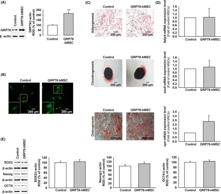
Objectives: The purpose of this study was to explore the effectiveness of concurrent GRP78 overexpression combined with Cripto on hMSC proliferation and migration both in vitro and in vivo. Specifically, we explored whether the treatment enhances effectiveness of hMSC transplantation in ischaemic tissue.

Materials and methods: Human MSCs obtained from human adipose tissue were cultured in α-minimum essential medium (Hyclone, Logan, UT, USA) supplemented with 10% (v/v) foetal bovine serum (Hyclone), 100 U mL-1 penicillin and 100 μg mL-1 streptomycin. Murine hindlimb ischaemic model was generated with 8-week-old male nude BALB/c mice (Biogenomics, Seoul, Korea) maintained under a 12-h light/dark cycle following the established protocol with minor modification. Cellular injection was performed no later than 3 hour after surgery. Lipofectamine transfection, single-cell cultivation assay, transwell assay, scratch wound-healing migration assay, immunohistochemistry and western blotting assays were performed.

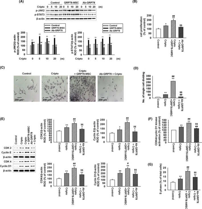
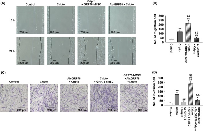
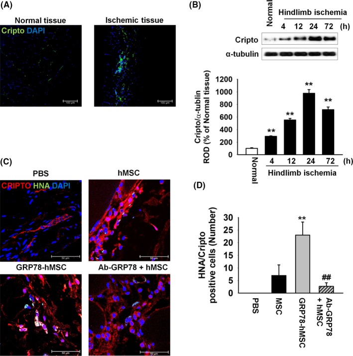
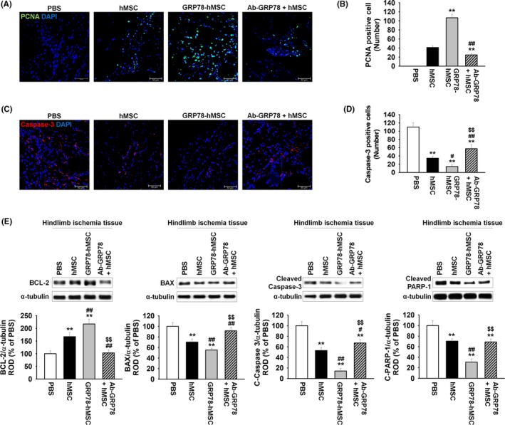
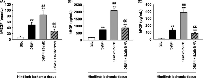
Results: Overexpression of GRP78 along with Cripto enhanced hMSC proliferation, migration and invasion. It increased interaction of surface GRP78 receptor with Cripto via JAK2/STAT3 pathway. We confirmed our proposed mechanism by showing that treatment with GRP78 antibody blocks the enhancement in vitro. In vivo, we observed that Cripto induced by the hypoxic environment in hindlimb ischaemic model interacts with the overexpressed GRP78 and increases hMSC proliferation, migration and invasion potentials as well as angiogenesis around transplanted ischaemic site via cytokine secretions.

Conclusions: These results demonstrate supporting evidences that GRP78-Cripto combination technique offers novel strategy to enhance MSC proliferation, migration and invasion potentials as well as angiogenesis around ischaemic site, ultimately facilitating MSC-based transplantation therapy in ischaemic conditions.

Keywords: Cripto; GRP78; angiogenesis; cell proliferation; invasion; mesenchymal stem cells.


Conflict of interest statement

The authors declare no conflict of interest.

Figures

Figure 1
Figure 1
Overexpression of GRP78 on human MSCs retains differentiation capabilities of stem cells. (A) Western blot analysis of GRP78 expression in hMSCs compared to that in overexpression GRP78‐hMSCs (GRP78‐hMSC). The right panel represents the expression levels of GRP78 that were normalized to β‐actin. Values represent mean ± SEM. **< .01 vs. Control. (B) GRP78‐hMSC was investigated by immunohistochemistry for GRP78 (Green). Scale bar = 200 μM. (C) hMSCs and GRP78‐hMSC were differentiated into adipocytes, osteocytes and chondrocytes, and examined by Oli red O, Safranin O, and alkaline phosphatase staining respectively. Scale bar = 200 μM. (D) mRNA expression of fabp4, sox9 and opn in hMSC and GRP78‐hMSC. (E) Western blot analysis of SOX2, Nanog and OCT4 expressions in GRP78‐hMSC and the control hMSC
Figure 2
Figure 2
Binding of GRP78 with Cripto enhances cellular proliferation via JAK2‐STAT3 pathway. (A) Western blot analysis of p‐JAK2 and p‐STAT3 expressions in hMSCs, GRP78‐hMSC and hMSC pre‐treated with antibody GRP78 (Ab‐GRP78) exposed to Cripto for 0, 5, 10 and 20 minutes. The down panel represents the phosphorylation levels of JAK2 and STAT‐3 in control to β‐actin. Values represent the mean ± SEM. **< .01 vs. control, ##< .01 vs. GRP78‐hMSC. (B) hMSC, GRP78‐hMSC, and Ab‐GRP78 were treated with or without Cripto for 24 hour. Images of cellular proliferation measurement were shown using MTT assay. Values represent the mean ± SEM. *P < .05 vs control, **< .01 vs. control, ##P < .01 vs treated Cripto, and $$P < .01 vs Cripto+GRP78‐hMSCs. (C) Image of single cell assay after 10 days in 96 well plate, stained with Giemsa stain. Scale bar = 100 μM. (D) The number of cells per field of view in each well of a 96 well plates is plotted (n = 3). Values represent the mean ± SEM. **P < .01 vs control, ##P < .01 vs treated Cripto, and $$P < .01 vs Cripto+GRP78‐hMSCs. (E) Western blot analysis of CDK 2, Cyclin E, CDK 4, and Cyclin D1 in hMSC, GRP78‐hMSC and Ab‐GRP78 were treated with or without Cripto for 24 hour. Values represent the mean ± SEM. *P < .05 vs control, **< .01 vs control, #P < .05 vs treated Cripto, ##P < .01 vs treated Cripto and $$P < .01 vs Cripto+GRP78‐hMSCs. (F) hMSC, GRP78‐hMSC and Ab‐GRP78 were treated with or without Cripto for 24 hour. Measurement of CDK4/cyclin D1 kinase activation concentration in hMSCs using ELISA (n = 3). Values represent the mean ± SEM. **< .01 vs untreated hMSCs. ##P < .01 vs treated Cripto, and $$P < .01 vs Cripto+GRP78‐hMSCs. (G) Images of flow cytometric analysis for PI staining to assess S phase populations in hMSC, GRP78‐hMSC and Ab‐GRP78 treated with Cripto. **< .01 vs untreated hMSCs. ##P < 0.01 vs treated Cripto and $$P < .01 vs Cripto+GRP78‐hMSCs
Figure 3
Figure 3
Overexpression of GRP78 on human MSCs increased migration and invasion through binding of surface GRP78 with Cripto. (A) Images of scratch wound‐healing migration assay of hMSCs, hMSCs with Cripto, GRP78+hMSC with Cripto and hMSCs with Cripto pre‐treated with Ab‐GRP78 for 0, and 24 hour. Scale bar = 100 μM. (B) The number of migrating cells is presented as the number of migrated cells per filed. Values represent mean ± SEM. **P < .01 vs control, ##P < .01 vs treated Cripto, and $$P < .01 vs Cripto+GRP78‐hMSC. (C) Invasion assay images of hMSCs, hMSCs with Cripto, hMSCs with Cripto pre‐treated with Ab‐GRP78, GRP78+hMSC with Cripto and GRP78+hMSCs with Cripto pre‐treated with Ab‐GRP78 for 24 hour. Scale bar = 100 μM. (D) Values represent the mean ± SEM. *P < .05 vs control, **P < .01 vs control, ##P < .01 vs treated Cripto, and $$P < .01 vs Ab‐GRP78‐hMSCs, &&P < .01 vs Cripto+GRP78‐hMSCs
Figure 4
Figure 4
In vivo, Cripto is overexpressed in hindlimb ischaemia and overexpression of GRP78 on human MSCs increased recruitment of Cripto to MSCs via GRP78 receptor of ischaemia injury site. (A) Images of immunohistochemistry results for Cripto (green) in the ischaemia injury tissue 3 days after the murine hindlimb ischaemia operation. Scale bar = 100 μm. (B) Western blot analysis showing the expression of Cripto in ischaemia injury tissue at post‐operative for 0, 4, 12, 24 and 72 hour. The down panel represents the expression levels of Cripto which were normalized to α‐tubulin. Values represent the mean ± SEM. **< .01 vs normal tissues. (C) At post‐operative of 3 days, immunohistochemistry assay for HNA (green) and Cripto (red) was performed in the ischaemia injury tissue of each group. Scale bar = 100 μm. (D) Existence of transplanted hMSCs were quantified based on the number of HNA and Cripto double positive cells. Values represent the mean ± SEM. **P < .01 vs. transplanted hMSCs, ##P < .01 vs transplanted overexpression GRP78 on hMSCs
Figure 5
Figure 5
Overexpression GRP78 on human MSCs decreased cell apoptosis in murine hindlimb ischaemia. (A) At post‐operative of 3 days, immunohistochemistry assay for PCNA (green) was performed in the ischaemia injury tissue of each group. Scale bar = 100 μm. (B) Cell proliferation was quantified as the number of PCNA positive cells. Values represent the mean ± SEM. **P < .01 vs transplanted hMSCs, ##P < .01 vs transplanted overexpression GRP78 on hMSCs. (C) At post‐operative of 3 days, immunohistochemistry assay for caspase‐3 (red) was performed in the ischaemia injury tissue of each group. Scale bar = 100 μm. (D) Cell apoptosis was quantified as the number of caspase‐3 positive cells. Values represent the mean ± SEM. **P < .01 vs injection of PBS, #P < .05, ##P < .01 vs transplanted hMSCs, and $$P < .01 vs transplanted overexpression GRP78 on hMSCs. (E) Western blot analysis showing the expression of BCL‐2, BAX, cleaved caspase‐3 and cleaved PARP‐1 in ischaemia injury tissue of each group at post‐operative for 3 days. The down panel represents the expression levels of GRP78 were normalized to α‐tubulin. Values represent the mean ± SEM. **P < .01 vs injection of PBS, #P < .05, ##P < .01 vs transplanted hMSCs, and $$P < .01 vs transplanted overexpression GRP78 on hMSCs
Figure 6
Figure 6
Overexpression GRP78 on human MSCs increased secretion of angiogenic cytokines in murine hindlimb ischaemia. (A) Measurements of human VEGF, HGF and FGF concentrations in ischaemia injury tissue of each group at post‐operative for 3 days using ELISA. Ischaemia injury tissue of each group at post‐operative for 3 days. Values represent the mean ± SEM. **P < .01 vs injection of PBS, ##P < .01 vs transplanted hMSCs, and $$P < .01 vs transplanted overexpression GRP78 on hMSCs

Similar articles

Cited by

References

    1. Nakagami H, Maeda K, Morishita R, et al. Novel autologous cell therapy in ischemic limb disease through growth factor secretion by cultured adipose tissue‐derived stromal cells. Arterioscler Thromb Vasc Biol. 2005;25:2542‐2547. - PubMed
    1. Vaananen HK. Mesenchymal stem cells. Ann Med. 2005;37:469‐479. - PubMed
    1. Phinney DG, Prockop DJ. Concise review: mesenchymal stem/multipotent stromal cells: the state of transdifferentiation and modes of tissue repair–current views. Stem Cells. 2007;25:2896‐2902. - PubMed
    1. Potapova IA, Gaudette GR, Brink PR, et al. Mesenchymal stem cells support migration, extracellular matrix invasion, proliferation, and survival of endothelial cells in vitro. Stem Cells. 2007;25:1761‐1768. - PubMed
    1. Andrades JA, Han B, Becerra J, Sorgente N, Hall FL, Nimni ME. A recombinant human TGF‐beta1 fusion protein with collagen‐binding domain promotes migration, growth, and differentiation of bone marrow mesenchymal cells. Exp Cell Res. 1999;250:485‐498. - PubMed

MeSH terms