. 2018 Apr;13(4):717-726.
doi: 10.4103/1673-5374.230300.

Intracerebroventricularly-administered 1-methyl-4-phenylpyridinium ion and brain-derived neurotrophic factor affect catecholaminergic nerve terminals and neurogenesis in the hippocampus, striatum and substantia nigra

Affiliations

Intracerebroventricularly-administered 1-methyl-4-phenylpyridinium ion and brain-derived neurotrophic factor affect catecholaminergic nerve terminals and neurogenesis in the hippocampus, striatum and substantia nigra

Jun-Fang Chen et al. Neural Regen Res. 2018 Apr.

Abstract

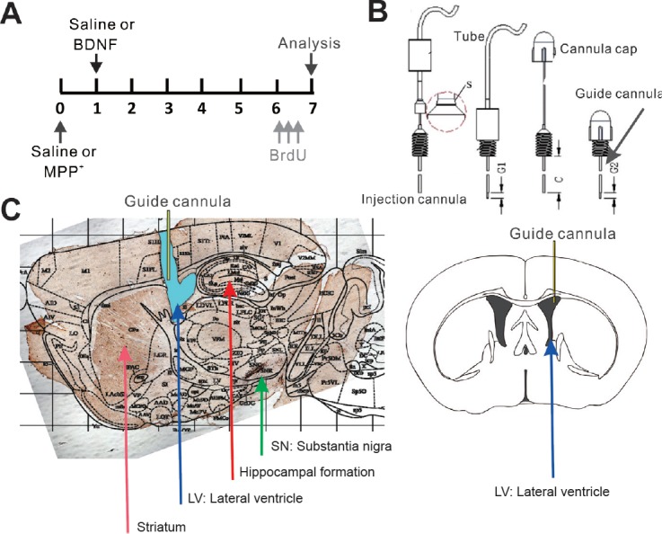
Parkinson's disease is a progressive neurological disease characterized by the degeneration of dopaminergic neurons in the substantia nigra. A highly similar pattern of neurodegeneration can be induced by 1-methyl-4-phenyl-1,2,3,6-tetrahydropyridine (MPTP) or 1-methyl-4-phenylpyridinium ion (MPP+), which cause the death of dopaminergic neurons. Administration of MPTP or MPP+ results in Parkinson's disease-like symptoms in rodents. However, it remains unclear whether intracerebroventricular MPP+ administration affects neurogenesis in the substantia nigra and subgranular zone or whether brain-derived neurotrophic factor alters the effects of MPP+. In this study, MPP+ (100 nmol) was intracerebroventricularly injected into mice to model Parkinson's disease. At 7 days after administration, the number of bromodeoxyuridine (BrdU)-positive cells in the subgranular zone of the hippocampal dentate gyrus increased, indicating enhanced neurogenesis. In contrast, a reduction in BrdU-positive cells was detected in the substantia nigra. Administration of brain-derived neurotrophic factor (100 ng) 1 day after MPP+ administration attenuated the effect of MPP+ in the subgranular zone and the substantia nigra. These findings reveal the complex interaction between neurotrophic factors and neurotoxins in the Parkinsonian model that result in distinct effects on the catecholaminergic system and on neurogenesis in different brain regions.

Keywords: MPTP; Parkinson's disease; brain-derived neurotrophic factor; dopaminergic fibers; hippocampus; intracerebroventricular infusion; nerve regeneration; neural regeneration; neurogenesis; norepinephrine; striatum; substantia nigra; tyrosine hydroxylase.


Conflict of interest statement

We declare that we have no conflict of interest

Figures

Figure 1
Figure 1
Illustration of the sequence of MPP+, BDNF and BrdU administration, the single-tube system for drug delivery, and the location of the cannula. (A) The temporal sequence of MPP+, BDNF and BrdU administration. Mice were randomly assigned to the four different groups, and single-tube delivery systems were implanted. To induce cell death of dopaminergic neurons, MPP+/saline was intracerebroventricularly administrated on day 0. For other experiments BDNF/saline was injected into the right lateral ventricle 1 day later. To assess neurogenesis, BrdU was intraperitoneally administrated 5 days later, three times (gray arrows). The mice were transcardially perfused 9 hours after the last intraperitoneal BrdU injection. (B) A single-tube delivery system is depicted. A cannula was placed into the guide cannula and connected to a microsyringe by a polyethylene tube, which was used to infuse the saline, MPP+ or BDNF at a rate of 0.3 µL/min. (C) An overlay of a TH-stained sagittal brain section with the corresponding regions outlined in the mouse brain atlas. The localization of the cannula (yellow) in the right LV (blue) is shown, as are the hippocampal formation, the striatum and the SN. To the right, the localization of the cannula in the LV is shown (coronal section). MPP+: 1-Methyl-4-phenylpyridinium ion; BDNF: brain-derived neurotrophic factor; BrdU: bromodeoxyuridine; TH: tyrosine hydroxylase; LV: lateral ventricle; SN: substantia nigra.
Figure 2
Figure 2
Distribution of TH-positive nerve terminals in the hippocampal formation. (A) Representative fluorescence images of anti-TH stained catecholaminergic fibers in the hippocampal formation: Arrows indicate representative examples of TH-positive fibers. Scale bar: 100 μm. (B) The number of fibers per 100 μm. The fiber distribution is diffuse and not uniform, with a significant high density within the DG. CA1, CA3 and DG subfields are indicated. **P < 0.01 (mean ± SEM, n = 3, Student's t-test). DG: Dentate gyrus; TH: tyrosine hydroxylase.
Figure 3
Figure 3
Intracerebroventricular administration of MPP+ significantly reduces TH levels in the hippocampal formation within 6 days. (A) Immunofluorescence images of TH-positive fibers 6 days after intracerebroventricular administration of saline or 100 nmol MPP+. (A1, A2) Magnified immunofluorescence images of the DG and CA1 region, respectively. Arrows indicate representative TH-positive fibers. Scale bar: 200 μm. (B) Changes in TH-positive fibers after saline (n = 4; white bar) or MPP+ (n = 4; black bar) administration. The area of TH-positive fibers (thresholded) within the CA1, CA3 and DG regions was normalized to the total area of the analyzed region. (C) Western blots for TH and β-actin. The bar graph (mean ± SEM) shows a reduction in the ratio of TH protein to actin after MPP+ administration (n = 3), compared with saline (n = 3). The results show that after intracerebroventricular infusion of MPP+, the dense network of dopaminergic fibers in the hippocampus was destroyed. **P < 0.01 (mean ± SEM, Student's t-test). CA1, CA3 and DG subfields are indicated. MPP+: 1-Methyl-4-phenylpyridinium ion; DG: dentate gyrus; TH: tyrosine hydroxylase.
Figure 4
Figure 4
Effects of BDNF and MPP+ on TH levels in striatal nerve terminals and the number of dopaminergic cells in the SN. (A, B) Photomicrographs of sagittal brain sections of the SN and the striatum in the saline, BDNF, MPP+ and MPP+/BDNF groups, respectively. TH signals are in dark gray to black. Scale bar: 300 μm. (C) Number of TH-positive cells in the SN in the different groups; n = 6 for the saline and BDNF groups, and n = 5 for the MPP+ and MPP+/BNDF groups. (D) TH signal ratio in the two parts of the striatum for the various conditions. To measure TH levels in the striatum, the mean intensity in an area of the striatum close to the ventricle was normalized to the mean intensity in a lateral region (striatum (ventricle)/striatum (lateral region)); n = 4 for the saline, BDNF and MPP+ groups, and n = 5 for the MPP+/BDNF group. Intracerebroventricular administration of MPP+ induced a significant reduction in TH-positive neurons in the SN and a change in TH levels in the striatum. BDNF did not attenuate the effect of MPP+ on the degeneration of SN neurons but affected striatal TH levels. *P < 0.05, **P < 0.01, ***P < 0.001 (mean ± SEM, one-way analysis of variance followed by Fisher's least significant difference test). SN: Substantia nigra; TH: tyrosine hydroxylase; BDNF: brain-derived neurotrophic factor; MPP+: 1-methyl-4-phenylpyridinium ion; ven: ventricle; lat: lateral region.
Figure 5
Figure 5
Confocal fluorescence images of BrdU-positive cells and their quantitative analysis in the SGZ, dentate gyrus and substantia nigra. (A) Fluorescence images of BrdU-positive cells in the SGZ of the dentate gyrus 6 days after administration of saline, BDNF, MPP+ or MPP+/BDNF. The white arrows indicate BrdU-positive cells, and the green arrow indicates the SGZ. (B) Quantification of BrdU-positive cells in the hippocampus after treatment with saline (n = 5), BDNF (n = 6), MPP+ (n = 4) or MPP+/BDNF (n = 4). (C) Fluorescence images of BrdU-positive cells (red) and TH-positive cells (green) in the substantia nigra. The white arrows indicate BrdU-positive cells. (D) Quantification of BrdU-positive cells in the substantia nigra after treatment with saline (n = 4), BDNF (n = 6), MPP+ (n = 4) or MPP+/BDNF (n = 3). MPP+ robustly increased BrdU-positive signals (neurogenesis) in the SGZ, and injection of BDNF blocked this effect of MPP+. BDNF did not prevent the MPP+-induced reduction in neurogenesis in the substantia nigra. *P < 0.05, **P < 0.01, ***P < 0.001 (mean ± SEM, one-way analysis of variance followed by Fisher's least significant difference test). Scale bar: 200 μm. TH: Tyrosine hydroxylase; BDNF: brain-derived neurotrophic factor; SGZ: subgranular zone; MPP+: 1-methyl-4-phenylpyridinium ion.

References

    1. Bellucci A, Antonini A, Pizzi M, Spano P. The end is the Beginning: Parkinson's disease in the light of brain imaging. Front Aging Neurosci. 2017;9:330. - PMC - PubMed
    1. Benmansour S, Deltheil T, Piotrowski J, Nicolas L, Reperant C, Gardier AM, Frazer A, David DJ. Influence of brain-derived neurotrophic factor (BDNF) on serotonin neurotransmission in the hippocampus of adult rodents. Eur J Pharmacol. 2008;587:90–98. - PubMed
    1. Binukumar BK, Pant HC. TFP5/TP5 peptide provides neuroprotection in the MPTP model of Parkinson's disease. Neural Regen Res. 2016;11:698–701. - PMC - PubMed
    1. Borodovitsyna O, Flamini M, Chandler D. Noradrenergic modulation of cognition in health and disease. Neural Plast. 2017 doi: 10.1155/2017/603147. - PMC - PubMed
    1. Braun DJ, Kalinin S, Feinstein DL. Conditional depletion of hippocampal brain-derived neurotrophic factor exacerbates neuropathology in a mouse model of Alzheimer's disease. ASN Neuro. 2017 doi: 10.1177/1759091417696161. - PMC - PubMed