. 2018 Aug 1;315(2):L313-L327.
doi: 10.1152/ajplung.00355.2017. Epub 2018 May 3.

Long-term culture and cloning of primary human bronchial basal cells that maintain multipotent differentiation capacity and CFTR channel function

Affiliations

Long-term culture and cloning of primary human bronchial basal cells that maintain multipotent differentiation capacity and CFTR channel function

Jennifer R Peters-Hall et al. Am J Physiol Lung Cell Mol Physiol. .

Abstract

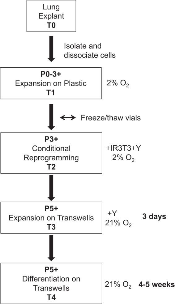
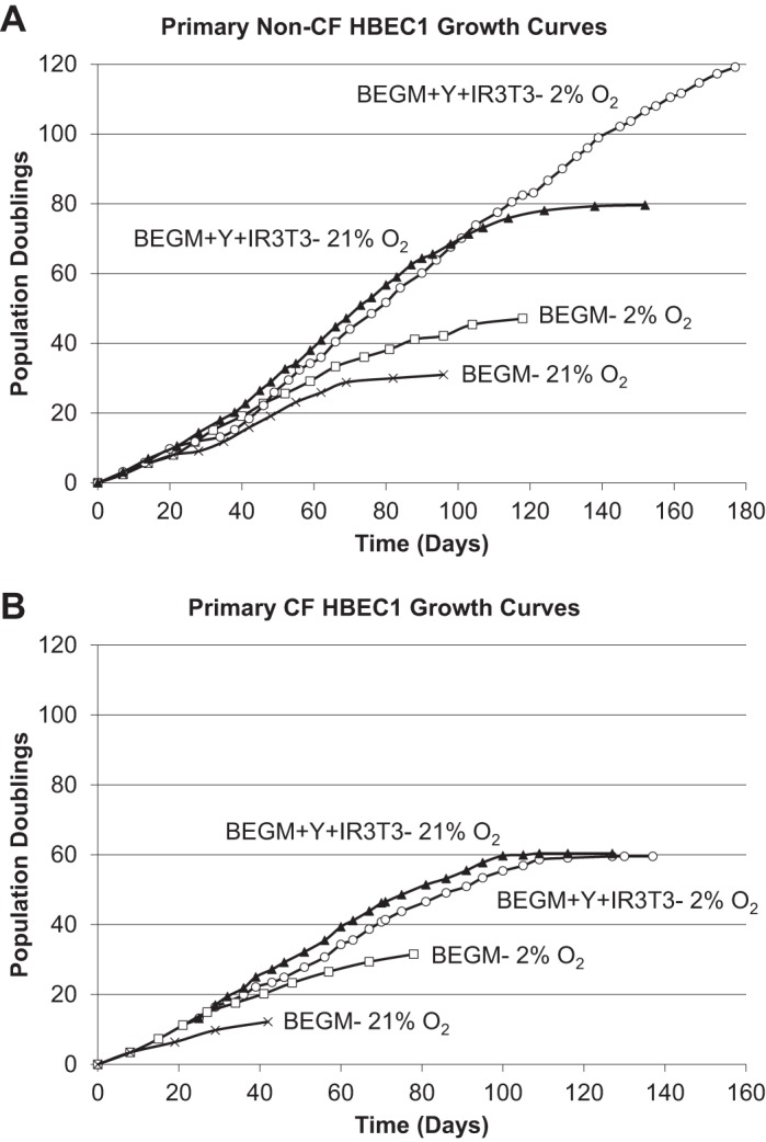
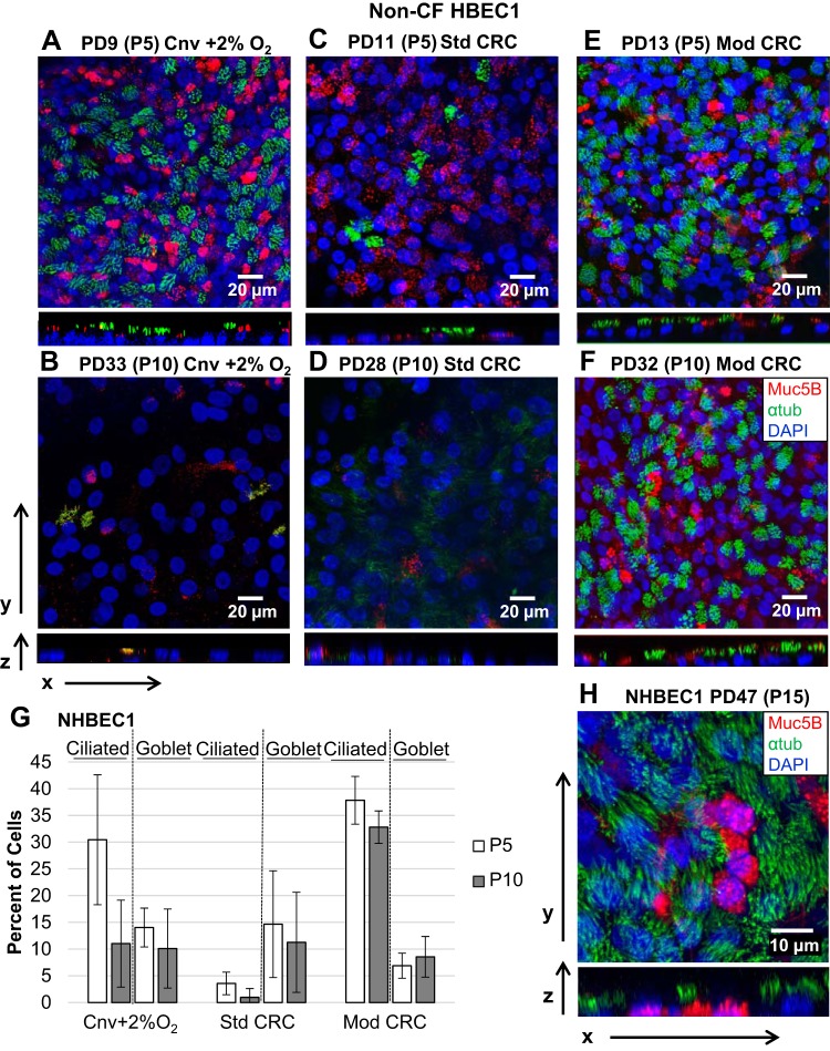
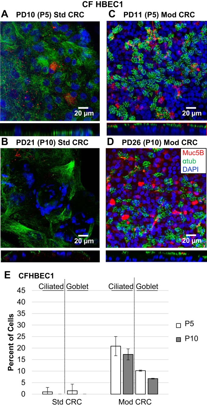
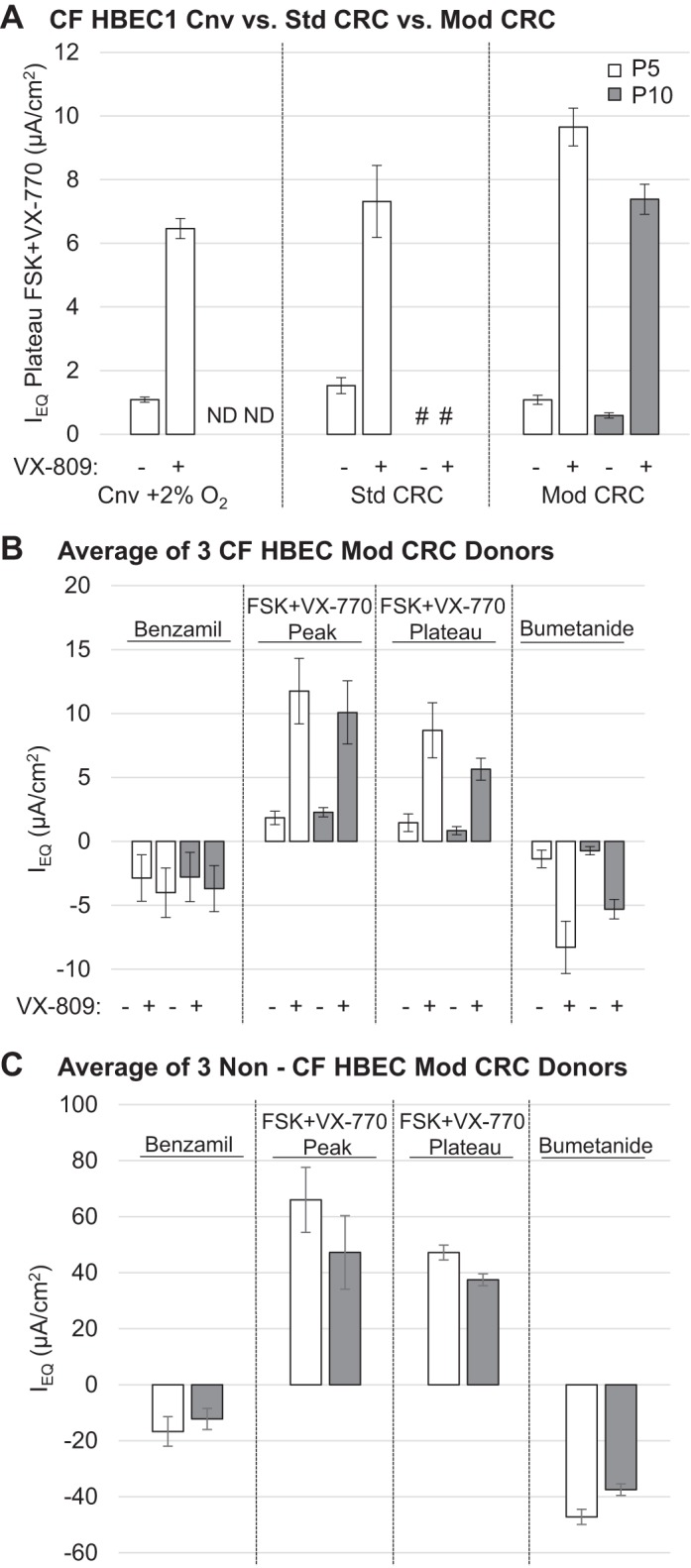
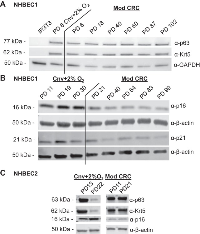
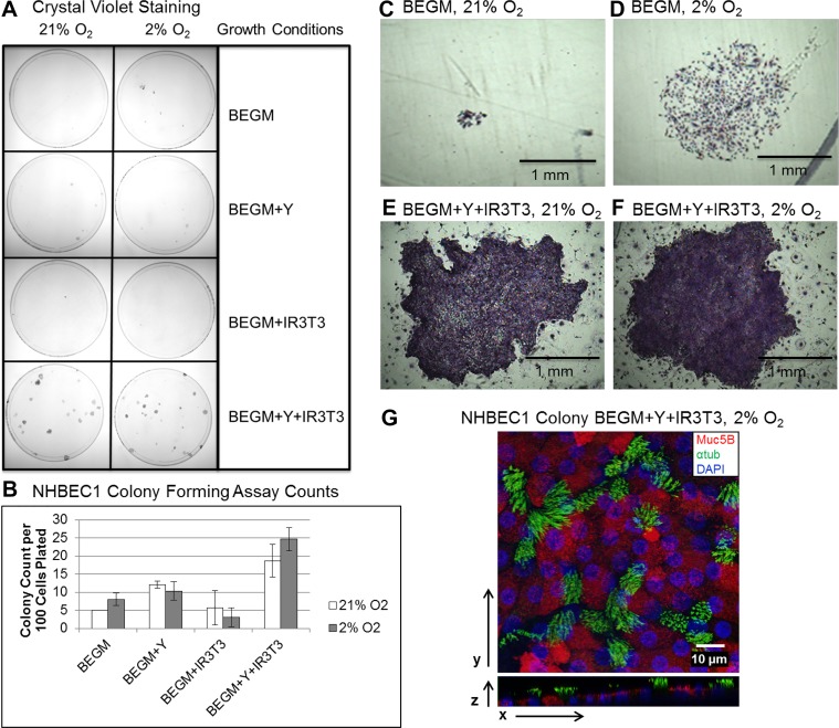
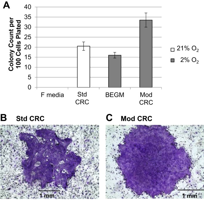
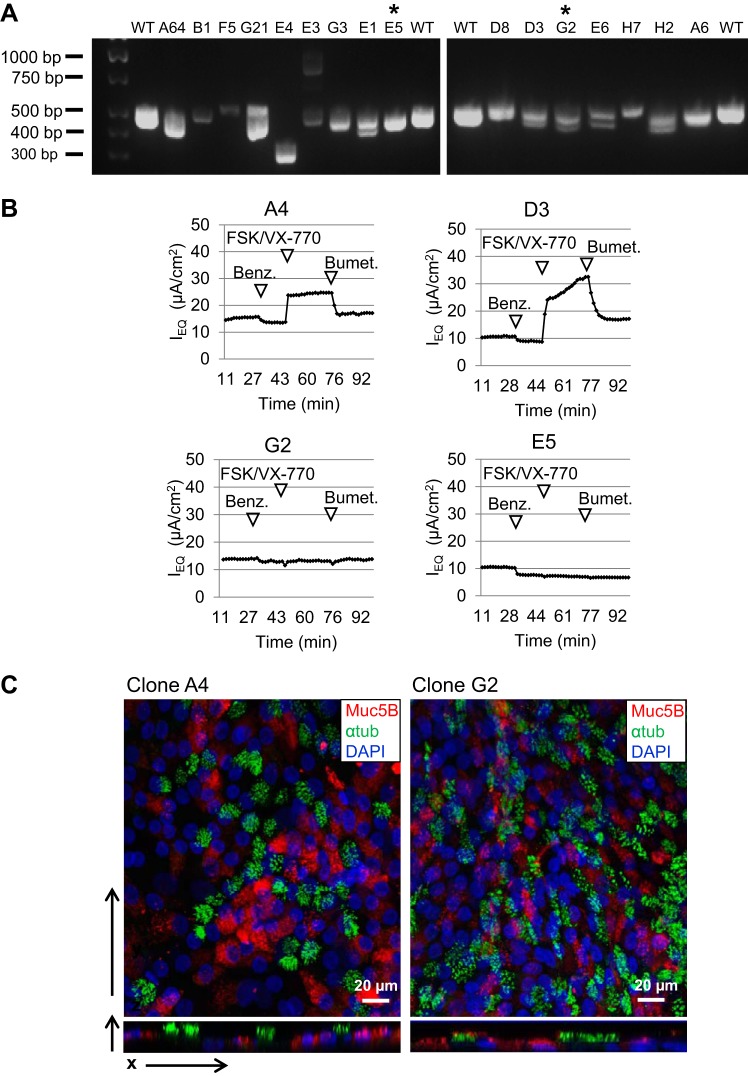
While primary cystic fibrosis (CF) and non-CF human bronchial epithelial basal cells (HBECs) accurately represent in vivo phenotypes, one barrier to their wider use has been a limited ability to clone and expand cells in sufficient numbers to produce rare genotypes using genome-editing tools. Recently, conditional reprogramming of cells (CRC) with a Rho-associated protein kinase (ROCK) inhibitor and culture on an irradiated fibroblast feeder layer resulted in extension of the life span of HBECs, but differentiation capacity and CF transmembrane conductance regulator (CFTR) function decreased as a function of passage. This report details modifications to the standard HBEC CRC protocol (Mod CRC), including the use of bronchial epithelial cell growth medium, instead of F medium, and 2% O2, instead of 21% O2, that extend HBEC life span while preserving multipotent differentiation capacity and CFTR function. Critically, Mod CRC conditions support clonal growth of primary HBECs from a single cell, and the resulting clonal HBEC population maintains multipotent differentiation capacity, including CFTR function, permitting gene editing of these cells. As a proof-of-concept, CRISPR/Cas9 genome editing and cloning were used to introduce insertions/deletions in CFTR exon 11. Mod CRC conditions overcome many barriers to the expanded use of HBECs for basic research and drug screens. Importantly, Mod CRC conditions support the creation of isogenic cell lines in which CFTR is mutant or wild-type in the same genetic background with no history of CF to enable determination of the primary defects of mutant CFTR.

Keywords: CRISPR; ROCK inhibitor; conditional reprogramming; cystic fibrosis; human bronchial epithelial cells.


Figures

Fig. 1.
Fig. 1.
Flowchart of human bronchial epithelial cell isolation, expansion, conditional reprogramming, and differentiation. T, time; P, passage; IR 3T3, freshly irradiated (30 Gy) 3T3 fibroblasts; Y,  Y-27632 (Rho-associated protein kinase inhibitor).
Fig. 2.
Fig. 2.
Growth curves of serially passaged human bronchial epithelial cells (HBECs) from a non-CF donor (NHBEC1) and a CF donor (CFHBEC1) in conventional bronchial epithelial growth medium (BEGM) and in modified conditionally reprogrammed (CRC) conditions with 21% atmospheric O2 or 2% O2. HBECs from non-CF (A) and CF (B) donors senesced after long-term culture in BEGM-21% O2 (×), and even in BEGM-2% O2 (□), while the same donors exhibited extended life span in CRC conditions with Rho-associated protein kinase (ROCK) inhibitor Y-27632 (Y), irradiated 3T3 fibroblast feeder layer (IR 3T3), and 21% O2 (▲) or 2% O2 (○). Growth of primary HBECs from the non-CF donor in CRC conditions (BEGM + Y + IR 3T3) was limited to 80 population doublings (PDs) in 21% O2, while 2% O2 (BEGM + Y + IR 3T3-2% O2) significantly extended the life span of HBECs from the non-CF HBEC donor beyond 100 PDs. However, the life span of HBECs from the CF donor was limited to 60 PDs in CRC conditions with either 21% or 2% O2.
Fig. 3.
Fig. 3.
Morphology of air-liquid interface cultures of human bronchial epithelial cells (HBECs) from a non-CF donor (NHBEC1) differentiated after conventional (Cnv) expansion (with 2% O2) or standard (Std) or modified (Mod) conditionally reprogrammed cell (CRC) expansion. A–F: representative air-liquid interface (ALI) cultures differentiated from HBECs from the non-CF donor at indicated population doublings (PDs) and passages (P) and growth conditions were immunostained for mucin 5B (Muc5B; red)-expressing goblet cells and acetylated α-tubulin (αtub; green)-expressing ciliated cells and counterstained with DAPI (blue). Confocal z-stack images were acquired and merged in the xy or zy plane. G: percentage of resulting ciliated and goblet cells at the indicated passage and growth condition for HBECs from the non-CF donor. Values are means ± SD; n = 3 images. H: multichannel fluorescence confocal microscopy shows that HBECs from the non-CF donor expanded in modified CRC conditions maintain the capacity to differentiate to a well-differentiated epithelium at the ALI as late as 47 PDs (passage 15), with abundant ciliated and goblet cells.
Fig. 4.
Fig. 4.
Morphology of air-liquid interface (ALI) cultures of HBECs from a CF donor (CFHBEC1) differentiated after standard (Std) or modified (Mod) conditionally reprogrammed cell (CRC) expansion. A–D: representative images of ALI cultures differentiated from HBECs from the CF donor at indicated population doubling (PD) and passage (P) and growth conditions were immunostained for mucin 5B (Muc5B; red)-expressing goblet and acetylated α-tubulin (αtub; green)-expressing ciliated cells and counterstained with DAPI (blue). Confocal z-stack images were acquired and merged in the xy or zy plane. E: percentage of resulting ciliated and goblet cells at the indicated passage and growth condition. Values are means ± SD; n = 3 images.
Fig. 5.
Fig. 5.
CFTR-dependent Cl secretion of air-liquid interface (ALI) cultures of human bronchial epithelial cells (HBECs) from CF (CFHBEC1) and non-CF (NHBEC1) donors previously expanded in conventional (Cnv), standard (Std), and modified (Mod) conditionally reprogrammed cell (CRC) conditions. A: HBECs from donor CFHBEC1 expanded in Cnv + 2% O2, Std, and Mod CRC conditions at passages 5 (P5) and 10 (P10) were differentiated at the ALI for 4−5 wk, and CFTR-dependent Cl secretion was analyzed by TECC-24 assay 48 h after treatment with 3 µM VX-809 (+) or DMSO (−). P5 = 10–13 population doublings (PDs); P10 = 20–26 PDs. Values are means ± SE; n = 3 experiments for Cnv and Mod CRC and 2 experiments for Std CRC because of unmeasurable results for transepithelial resistance (Rt) and equivalent current (Ieq) in the 3rd experiment, 6–8 replicates. #No detectable Rt/Ieq. ND, not determined. B: average Ieq of ALI cultures of HBECs from donors CFHBEC1, CFHBEC2, and CFHBEC3 previously expanded in Mod CRC conditions in response to benzamil, forskolin (FSK) + VX-770, or bumetanide 48 h after treatment with 3 µM VX-809 (+) or DMSO (−). P5 = 10–13 PDs; P10 = 20–26 PDs. Values are means ± SE for 1 experiment, 3 donors, 5–6 replicates. C: average Ieq of ALI cultures of HBECs from donors NHBEC1, NHBEC2, and NHBEC3 previously expanded in Mod CRC conditions in response to benzamil, FSK + VX-770, or bumetanide. P5 = 9–13 PDs; P10 = 27–29 PDs. Values are means ± SE for 1 experiment, 3 donors, 5–6 replicates.
Fig. 6.
Fig. 6.
Expression of basal progenitor human bronchial epithelial cell (HBEC) markers is preserved at late passage, while senescence-associated proteins are suppressed in modified (Mod) conditionally reprogrammed cell (CRC) HBECs from a non-CF donor (NHBEC1). A and B: Western blot analysis of p63, keratin 5 (Krt5), and p16 or p21, as well as GAPDH and β-actin loading controls, for protein cell lysates from HBECs from donor NHBEC1 grown over time in conventional (Cnv) + 2% O2 or Mod CRC conditions. IR3T3, irradiated 3T3 fibroblasts. PD, population doubling. C: Western blot analysis of p63, keratin 5, and p16 for protein cell lysates from HBECs from donor NHBEC2 grown for 5 or 8 passages in Cnv + 2% O2 or Mod CRC conditions.
Fig. 7.
Fig. 7.
Human bronchial epithelial cells (HBECs) from a non-CF donor (NHBEC1) seeded at clonal density require Y-27632 (Y), irradiated 3T3 fibroblasts (IR3T3), and 2% O2 (Mod CRC conditions) to form the largest and most numerous clones with normal epithelial morphology. A: HBECs from donor NHBEC1 at 20 population doublings (PDs) in modified (Mod) CRC conditions were seeded at clonal density in 10-cm dishes in the 8 different HBEC growth conditions and allowed to grow for 10 days before fixation and staining with crystal violet. Representative dishes were imaged. BEGM, bronchial epithelial cell growth medium. B: average colony counts of 3 dishes for each of the conditions in A. Values are means ± SD. C–F: representative crystal violet-stained colonies for BEGM and BEGM + Y + IR 3T3 conditions with 21% or 2% O2. G: multichannel fluorescence confocal microscopy of a HBEC clone from donor NHBEC1 under Mod CRC conditions differentiated at the air-liquid interface and immunostained for mucin 5B (Muc5B)-expressing goblet cells (red) or acetylated α-tubulin (αtub)-expressing ciliated cells (green).
Fig. 8.
Fig. 8.
Modified (Mod) conditionally reprogrammed cell (CRC) conditions support enhanced colony growth of human bronchial epithelial cells (HBECs) from a non-CF donor (NHBEC1) compared with standard (Std) CRC conditions. HBECs from donor NHBEC1 at 20 population doublings (PDs) were seeded at clonal density in 10-cm dishes in Std CRC or Mod CRC conditions or in F medium + 5% FBS (F medium) or bronchial epithelial cell growth medium (BEGM) alone as controls. Colonies were grown for 10 days before fixation and staining with crystal violet. A: average colony counts for 2 dishes for each condition. No colonies grew in F medium control conditions. Values are means ± SD. B and C: representative crystal violet-stained colonies of HBECs from donor NHBEC1 in Std and Mod conditions.
Fig. 9.
Fig. 9.
Modified conditionally reprogrammed cell (CRC) conditions enable efficient cloning and editing of CFTR by CRISPR/Cas9 in primary human bronchial epithelial cells (HBECs) from a non-CF donor (NHBEC1). A: for each of 16 clones, the region of CFTR exon 11 surrounding the CRISPR/Cas9 cut site was PCR-amplified from genomic DNA and separated by electrophoresis on two 2% gels, along with PCR product of genomic DNA from unedited HBECs with wild-type (WT) CFTR. *Clones G2 and E5 were selected for sequencing and further characterization. B: representative traces of 6–9 replicates from TECC-24 assay showed CFTR-dependent Cl secretion in clones A4 and D3, but not in clones G2 and E5. Ieq, equivalent current; FSK, forskolin; Benz, benzamil; Bumet, bumetanide. C: multichannel fluorescence confocal microscopy shows that WT and mutant CFTR clones maintain the capacity to form a well-differentiated epithelium at the air-liquid interface, with abundant ciliated cells expressing acetylated α-tubulin (αtub, green) and goblet cells expressing mucin 5B (Muc5B, red).

Similar articles

Cited by

References

    1. Boat TF, Welsh MJ, Beaudet AL. Cystic Fibrosis. New York: McGraw-Hill, 1989.
    1. Chapman S, Liu X, Meyers C, Schlegel R, McBride AA. Human keratinocytes are efficiently immortalized by a Rho kinase inhibitor. J Clin Invest 120: 2619–2626, 2010. doi:10.1172/JCI42297. - DOI - PMC - PubMed
    1. Coppé JP, Desprez PY, Krtolica A, Campisi J. The senescence-associated secretory phenotype: the dark side of tumor suppression. Annu Rev Pathol 5: 99–118, 2010. doi:10.1146/annurev-pathol-121808-102144. - DOI - PMC - PubMed
    1. Crane AM, Kramer P, Bui JH, Chung WJ, Li XS, Gonzalez-Garay ML, Hawkins F, Liao W, Mora D, Choi S, Wang J, Sun HC, Paschon DE, Guschin DY, Gregory PD, Kotton DN, Holmes MC, Sorscher EJ, Davis BR. Targeted correction and restored function of the CFTR gene in cystic fibrosis induced pluripotent stem cells. Stem Cell Reports 4: 569–577, 2015. doi:10.1016/j.stemcr.2015.02.005. - DOI - PMC - PubMed
    1. da Paula AC, Ramalho AS, Farinha CM, Cheung J, Maurisse R, Gruenert DC, Ousingsawat J, Kunzelmann K, Amaral MD. Characterization of novel airway submucosal gland cell models for cystic fibrosis studies. Cell Physiol Biochem 15: 251–262, 2005. doi:10.1159/000087235. - DOI - PubMed

Publication types

MeSH terms

Substances