

Some Isolated Cardiac Malformations Can Be Related to Laterality Defects
- PMID: 29724030
- PMCID: PMC6023464
- DOI: 10.3390/jcdd5020024
Some Isolated Cardiac Malformations Can Be Related to Laterality Defects
Abstract
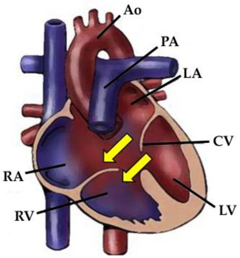
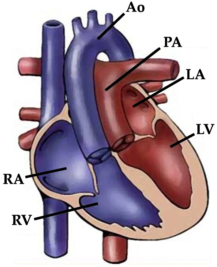
Human beings are characterized by a left⁻right asymmetric arrangement of their internal organs, and the heart is the first organ to break symmetry in the developing embryo. Aberrations in normal left⁻right axis determination during embryogenesis lead to a wide spectrum of abnormal internal laterality phenotypes, including situs inversus and heterotaxy. In more than 90% of instances, the latter condition is accompanied by complex and severe cardiovascular malformations. Atrioventricular canal defect and transposition of the great arteries—which are particularly frequent in the setting of heterotaxy—are commonly found in situs solitus with or without genetic syndromes. Here, we review current data on morphogenesis of the heart in human beings and animal models, familial recurrence, and upstream genetic pathways of left⁻right determination in order to highlight how some isolated congenital heart diseases, very common in heterotaxy, even in the setting of situs solitus, may actually be considered in the pathogenetic field of laterality defects.
Keywords: atrioventricular canal defect; congenital heart disease; genetics; heterotaxy; transposition of the great arteries.
Conflict of interest statement
The authors declare no conflict of interest.
Figures
References
-
- Nonaka S., Tanaka Y., Okada Y., Takeda S., Harada A., Kanai Y., Kido M., Hirokawa N. Randomization of left-right asymmetry due to loss of nodal cilia generating leftward flow of extraembryonic fluid in mice lacking KIF3B motor protein. Cell. 1998;95:829–837. doi: 10.1016/S0092-8674(00)81705-5. - DOI - PubMed
-
- Aranburu A., Piano Mortari E., Baban A., Giorda E., Cascioli S., Marcellini V., Scarsella M., Ceccarelli S., Corbelli S., Cantarutti N., et al. Human B-cell memory is shaped by age- and tissue-specific T-independent and GC-dependent events. Eur. J. Immunol. 2017;47:327–344. doi: 10.1002/eji.201646642. - DOI - PubMed
Publication types
LinkOut - more resources
Full Text Sources
Other Literature Sources
