. 2018 Oct;24(19-20):1492-1503.
doi: 10.1089/ten.TEA.2017.0492. Epub 2018 Aug 20.

Assembly of Tissue-Engineered Blood Vessels with Spatially Controlled Heterogeneities

Affiliations

Assembly of Tissue-Engineered Blood Vessels with Spatially Controlled Heterogeneities

Hannah A Strobel et al. Tissue Eng Part A. 2018 Oct.

Abstract

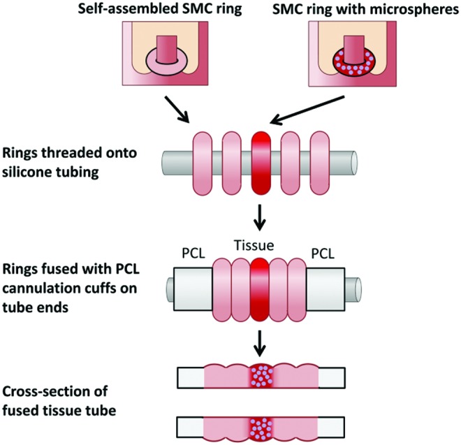
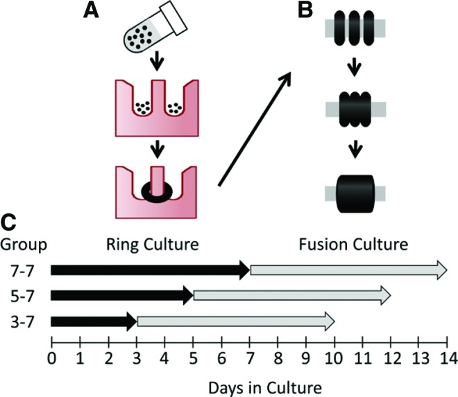
Tissue-engineered human blood vessels may enable in vitro disease modeling and drug screening to accelerate advances in vascular medicine. Existing methods for tissue-engineered blood vessel (TEBV) fabrication create homogenous tubes not conducive to modeling the focal pathologies characteristic of certain vascular diseases. We developed a system for generating self-assembled human smooth muscle cell (SMC) ring units, which were fused together into TEBVs. The goal of this study was to assess the feasibility of modular assembly and fusion of ring building units to fabricate spatially controlled, heterogeneous tissue tubes. We first aimed to enhance fusion and reduce total culture time, and determined that reducing ring preculture duration improved tube fusion. Next, we incorporated electrospun polymer ring units onto tube ends as reinforced extensions, which allowed us to cannulate tubes after only 7 days of fusion, and culture tubes with luminal flow in a custom bioreactor. To create focal heterogeneities, we incorporated gelatin microspheres into select ring units during self-assembly, and fused these rings between ring units without microspheres. Cells within rings maintained their spatial position along tissue tubes after fusion. Because tubes fabricated from primary SMCs did not express contractile proteins, we also fabricated tubes from human mesenchymal stem cells, which expressed smooth muscle alpha actin and SM22-α. This work describes a platform approach for creating modular TEBVs with spatially defined structural heterogeneities, which may ultimately be applied to mimic focal diseases such as intimal hyperplasia or aneurysm.

Keywords: bioreactor; electrospun cannulation cuff; gelatin microspheres; modular tissue engineering; tissue-engineered blood vessel; vascular disease model.


Conflict of interest statement

The authors have no competing financial interests.

Figures

<b>FIG. 1.</b>
FIG. 1.
Fabrication of modular tissue tubes with focal heterogeneities. Rings with incorporated microspheres are fused between rings without microspheres, with PCL cuffs on either end. The resulting construct is a fused tissue tube with focal region of microsphere incorporation. PCL, polycaprolactone. Color images available online at www.liebertpub.com/tea
<b>FIG. 2.</b>
FIG. 2.
Schematic of tube fabrication process, and tissue tube culture experimental groups for the ring preculture duration experiment. Rings are formed by seeding SMCs into ring-shaped agarose molds, where cells aggregate around 2 mm diameter posts and form rings in less than 24 h (A). Rings are then removed from molds and threaded onto silicone tubing, where they are pushed together and cultured for 7 additional days to allow fusion (B). To test the effects of varying ring culture duration, rings were cultured for 3, 5, or 7 days (“ring culture”), followed by 7 days of “fusion culture” for all groups (C). Groups are labeled as follows: days in ring culture–days in fusion culture (ex. Group 3–7 = 3 days in ring culture followed by 7 days in fusion culture). Black dots = SMCs. SMC, smooth muscle cell. Color images available online at www.liebertpub.com/tea
<b>FIG. 3.</b>
FIG. 3.
Fusion kinetics of human SMC rings. Three human SMC rings were threaded onto silicone tubing mandrels (A). The angle between rings (formula image
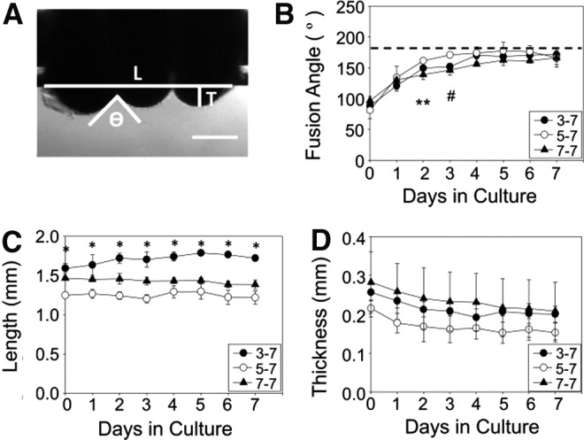
), tube length (L), and thickness (T) was measured for each sample on each day of culture (A). Fusion angles (B), tube length (C), and thickness (D) as a function of time for tubes fabricated from rings cultured for 3 (3–7), 5 (5–7), or 7 (7–7) days before 7 days of fusion culture. N = 3 tubes per group. Data points are mean ± SD. #p < 0.05 for 5–7 versus 3–7 and 7–7, ** p < 0.05 for 5–7 versus 7–7, *p < 0.05. Scale = 0.5 mm.
<b>FIG. 4.</b>
FIG. 4.
Histological assessment of human SMC tubes. H&E-stained tissue tubes comprised rings precultured for 3 (A–C), 5 (D–F), or 7 (G–I) days before fusion. Low magnification longitudinal sections shown in (A, D, G). Higher magnification views show one fusion point at the outer surfaces (solid box; B, E, H) and at the inner surfaces (dashed box; C, F, I) of the tissue tubes. Lumen on bottom, scale bars = 250 μm (low magnification) or 100 μm (high magnification). Images representative from n = 3 samples/group. Sectioning schematic shown in lower right. Color images available online at www.liebertpub.com/tea
<b>FIG. 5.</b>
FIG. 5.
Spatial position of rings during fusion. Human aortic SMCs were preloaded with red or green CellTracker dye before ring seeding. Rings with alternating dyes were then stacked and allowed to fuse for 7 days (A). Tubes were then sectioned and stained with Hoechst dye. Red = CellTracker Red (B), green = CellTracker Green (C), and blue = nuclei (D). Merged image shown in (E). Scale = 1 mm (A) or 100 μm (B–E). Lumen on bottom (B–E). Images representative from n = 3 samples. Color images available online at www.liebertpub.com/tea
<b>FIG. 6.</b>
FIG. 6.
Cell proliferation during fusion. Human aortic SMCs were preloaded with red CellTracker dye before ring seeding. Rings were allowed to fuse for 1 (3-1 Tube) or 2 (3-2 Tube) days. Tubes were then sectioned and stained for Ki67 to examine proliferation. Green = Ki67, red = CellTracker Red, blue = nuclei. Scale = 100 μm. Images representative of n = 2 samples. Color images available online at www.liebertpub.com/tea
<b>FIG. 7.</b>
FIG. 7.
PCL cannulation cuff incorporation for bioreactor culture. Electrospun PCL cuffs were threaded onto silicone tubing and pushed into contact with cell rings at each end of the tube. Tubes were cultured for 7 days on silicone mandrels (A) to achieve ring fusion, and then mounted onto the cannulas in the chamber of a custom luminal flow bioreactor (B). Image of bioreactor with SMC tube is shown in (C), and a schematic of the medium flow loop is shown in (D). Scale = 1 cm. Color images available online at www.liebertpub.com/tea
<b>FIG. 8.</b>
FIG. 8.
Histological images of tubes cultured in a luminal flow bioreactor. Hematoxylin and eosin stain of longitudinal section of tissue tubes cultured as rings for 3 days, fused as tubes for 7 days, and then cultured on silicone mandrels in static conditions (A, B) or with ∼12 dyne/cm2 shear stress (C, D) for an additional 7 days. Lumen at bottom of image. Scale = 100 μm. Color images available online at www.liebertpub.com/tea
<b>FIG. 9.</b>
FIG. 9.
Matrix deposition in fused tissue tubes. Longitudinal sections of tubes cultured in static conditions for 14 days (A, B), or in static conditions for 7 days followed by 7 days of dynamic culture with ∼12 dyne/cm2 of applied shear (C, D). Picrosirius red fast green stain shown in (A, C; red = collagen, green = counterstain), orcein stain shown in (B, D; dark pink = elastic fibers, purple = nuclei). Lumen on bottom of image. Scale = 100 μm. Color images available online at www.liebertpub.com/tea
<b>FIG. 10.</b>
FIG. 10.
Human SMC tube with spatial heterogeneity. Human aortic SMC rings were either loaded with gelatin microspheres and red CellTracker dye, or without microspheres or dye. Rings with microspheres were placed in the central region of the tube, between outer regions without microspheres. Photograph of fused tissue tube shown in (A). PCL cuffs on either end reinforce the tube ends to aid in handling and cannulation. Low (B) and high (C) magnification H&E images of the vessel wall show that rings appear well fused, and microspheres maintain their spatial position in the center. Lumen on bottom of image (B, C). Scale in mm (A) or 100 μm (B, C). Images representative of two samples. Color images available online at www.liebertpub.com/tea
<b>FIG. 11.</b>
FIG. 11.
Contractile protein expression in fused hMSC tubes. Tubes were fabricated from 3-day-old hMSC rings, which were allowed to fuse for 7 days in static conditions. Green = SMA (A), SM22-α (B), or calponin (C), blue = nuclei. Lumen on bottom of image. Scale = 100 μm. hMSC, human mesenchymal stem cell. Color images available online at www.liebertpub.com/tea
<b>FIG. 12.</b>
FIG. 12.
Alignment of hMSCs within hMSC tubes. Radial cross-section of hMSC tube after 4 days of fusion. Hematoxylin and eosin stain. Lumen on bottom of image. Scale = 100 μm. Color images available online at www.liebertpub.com/tea

References

    1. Go A.S., Mozaffarian D., Roger V.L., et al. . Heart disease and stroke statistics—2014 update: a report from the American Heart Association. Circulation 129, e28, 2014 - PMC - PubMed
    1. Stegemann J.P., and Nerem R.M. Altered response of vascular smooth muscle cells to exogenous biochemical stimulation in two- and three-dimensional culture. Exp Cell Res 283, 146, 2003 - PubMed
    1. Alexander J.H., Hafley G., Harrington R.A., et al. . Efficacy and safety of edifoligide, an E2F transcription factor decoy, for prevention of vein graft failure following coronary artery bypass graft surgery: PREVENT IV: A randomized controlled trial. JAMA 294, 2446, 2005 - PubMed
    1. Kim F.Y., Marhefka G., Ruggiero N.J., Adams S., and Whellan D.J. Saphenous vein graft disease: review of pathophysiology, prevention, and treatment. Cardiol Rev 21, 101, 2013 - PubMed
    1. Wystrychowski W., McAllister T.N., Zagalski K., Dusserre N., Cierpka L., and L'Heureux N. First human use of an allogeneic tissue-engineered vascular graft for hemodialysis access. J Vasc Surg 60, 1353, 2014 - PubMed

Publication types

LinkOut - more resources