. 2018 Jul;38(7):1528-1536.
doi: 10.1161/ATVBAHA.118.310906. Epub 2018 May 3.

Establishing the Transient Mass Balance of Thrombosis: From Tissue Factor to Thrombin to Fibrin Under Venous Flow

Affiliations

Establishing the Transient Mass Balance of Thrombosis: From Tissue Factor to Thrombin to Fibrin Under Venous Flow

Shu Zhu et al. Arterioscler Thromb Vasc Biol. 2018 Jul.

Abstract

Objective: We investigated the coregulation of thrombin and fibrin as blood flows over a procoagulant surface.

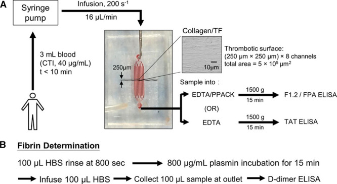
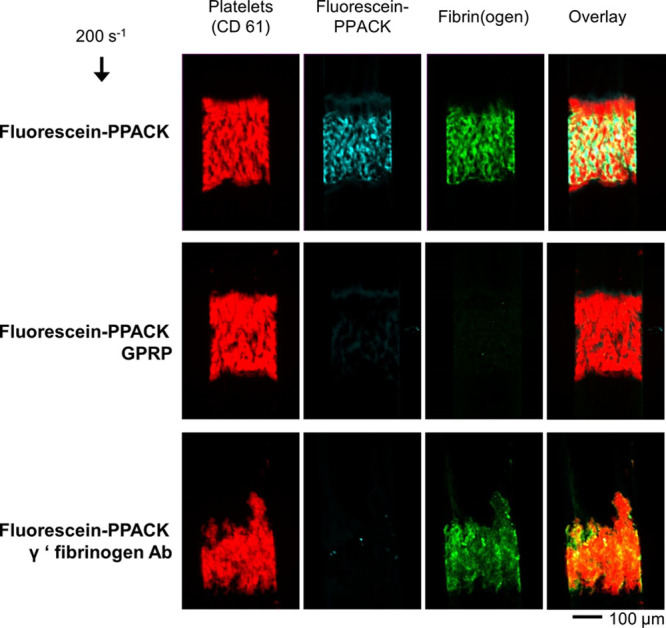
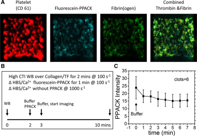
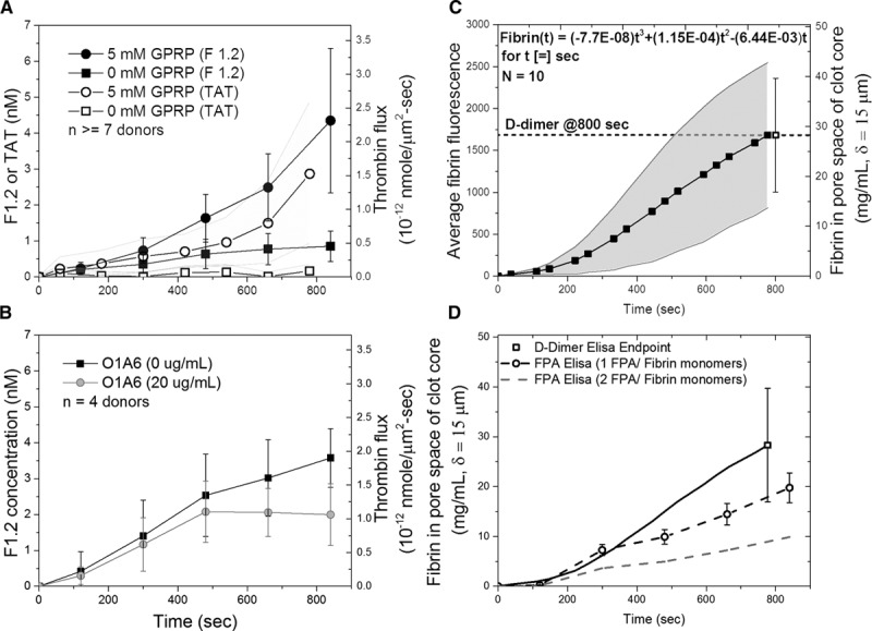
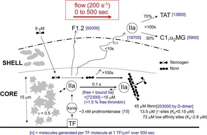
Approach and results: Using microfluidic perfusion of factor XIIa-inhibited human whole blood (200 s-1 wall shear rate) over a 250-μm long patch of collagen/TF (tissue factor; ≈1 molecule per μm2) and immunoassays of the effluent for F1.2 (prothrombin fragment 1.2), TAT (thrombin-antithrombin complex), and D-dimer (post-end point plasmin digest), we sought to establish the transient mass balance for clotting under venous flow. F1.2 (but almost no free thrombin detected via TAT assay) continually eluted from clots when fibrin was allowed to form. Low-dose fluorescein-Phe-Pro-Arg-chloromethylketone stained fibrin-bound thrombin-a staining ablated by anti-γ'-fibrinogen or the fibrin inhibitor glypro-arg-pro but highly resistant to 7-minute buffer rinse, demonstrating tight binding of thrombin to γ'-fibrin. With fibrin polymerizing for 500 seconds, 92 000 thrombin molecules and 203 000 clot-associated fibrin monomer equivalents were generated per TF molecule (or per μm2). Fibrin reached 15 mg/mL in the pore space (porosity ≈0.5) of a 15-μm-thick thrombus core by 500 seconds and 30 mg/mL by 800 seconds. For a known rate of ≈60 FPA (fibrinopeptide-A) per thrombin per second, each thrombin molecule generated only 3 fibrin monomer equivalents during 500 seconds, indicating an intraclot thrombin half-life of ≈70 ms, much shorter than its diffusional escape time (≈10 seconds). By 800 seconds, gly-pro-arg-pro allowed 4-fold more F1.2 generation, consistent with gly-pro-arg-pro ablating fibrin's antithrombin-I activity and facilitating thrombin-mediated FXIa activation.

Conclusions: Under flow, fibrinogen continually penetrates the clot, and γ'-fibrin regulates thrombin.

Keywords: blood; fibrin; hemodynamics; hemostasis; platelets; thrombosis.


Figures

Figure 1.
Figure 1.
Experimental protocol to perfuse whole blood over discrete 250-μm-long collagen/TF (tissue factor) surfaces while measuring dynamic platelet and fibrin accumulation by fluorescence microscopy and detecting F1.2 (prothrombin fragment 1.2) and TAT (thrombin–antithrombin complex) in the effluent using immunoassays (A). In some experiments, the clot was rinsed in situ and subjected to plasmin digestion to release D-dimer for subsequent immunoassay (B). CTI indicates corn trypsin inhibitor; FPA, fibrinopeptide-A; HBS, HEPES buffered saline; and PPACK, Phe-Pro-Arg-chloromethylketone.
Figure 2.
Figure 2.
Accumulation of fluorescent platelets and fibrin(ogen) on collagen/TF (tissue factor) in the presence of low-dose fluorescein-Phe-Pro-Arg-chloromethylketone (PPACK) to stain for the thrombin-active site under control conditions (top) or GPRP (gly-pro-arg-pro) to block fibrin (middle) or anti-γ′-fibrinogen antibody (bottom).
Figure 3.
Figure 3.
Washout experiment of fluorescein-Phe-Pro-Arg-chloromethylketone (PPACK)–labeled thrombin (A). Clots were formed for 2 min, stained with fluorescein-PPACK for 1 min, and then buffer washed for 10 min (B) to follow the elution of thrombin from the clot (C). Results are expressed as mean±SD (n=6). All the values are significantly different from zero (P<0.001) demonstrating essentially no washout of thrombin. CTI indicates corn trypsin inhibitor; HBS, HEPES buffered saline; TF, tissue factor; and WB, whole blood.
Figure 4.
Figure 4.
Comparison of F1.2 (prothrombin fragment 1.2) and TAT (thrombin–antithrombin complex) generation by whole blood clotting under venous flow conditions in the presence or absence of GPRP (gly-pro-arg-pro; A) or anti-FXIa antibody O1A6 (B). Alternatively, clots were formed with fluorescent fibrinogen tracer and then subjected to end point plasmin digest for assay of D-dimer (C). FPA (fibrinopeptide-A) generation was measured from 0 to 800 s. The result from independently measured D-dimer assay (dark line) was compared with assuming 1 FPA/fibrin monomer (dark dashed line) and 2 FPA/fibrin monomer (gray dashed line; D). Results are expressed as mean±SD.
Figure 5.
Figure 5.
Transient mass balance for production in the thrombus core (15-μm thick, porosity ≈0.5) of thrombin and fibrin during 500 s of venous thrombosis on collagen/TF (tissue factor). During 500 s, the copy number [blue] per unit area of 1 μm2 (at 1 TF per μm2) in a pore volume of 7.5 μm3 was calculated for total thrombin generation (based on F1.2 [prothrombin fragment 1.2] assay), amount of thrombin escape in the presence of fibrin (based on TAT [thrombin–antithrombin complex] assay), deposited fibrin (based on D-dimer assay), and active prothrombinase (based on known kinetic constants).
Figure 6.
Figure 6.
Calculation of free thrombin within the pore volume of the thrombus core at 500 s based on a total thrombin concentration of 15 μM and total fibrin concentration of 45 μM. The free thrombin concentration is shown for weak sites (E-domain) only with Kd of 2.8, 5 μM (dotted lines) or for weak sites in combination with the γ′-site with the γ′-Kd varied from 1 to 200 nmol/L (solid lines for weak Kd of 1 μM [circle], 2.8 μM [triangle], or 5 μM [square]). The weak and γ′-site concentrations were calculated to be 72 and 13.5 μM, respectively.

References

    1. Stalker TJ, Traxler EA, Wu J, Wannemacher KM, Cermignano SL, Voronov R, Diamond SL, Brass LF. Hierarchical organization in the hemostatic response and its relationship to the platelet-signaling network. Blood. 2013;121:1875–1885. doi: 10.1182/blood-2012-09-457739. - PMC - PubMed
    1. Welsh JD, Poventud-Fuentes I, Sampietro S, Diamond SL, Stalker TJ, Brass LF. Hierarchical organization of the hemostatic response to penetrating injuries in the mouse macrovasculature. J Thromb Haemost. 2017;15:526–537. doi: 10.1111/jth.13600. - PMC - PubMed
    1. Welsh JD, Colace TV, Muthard RW, Stalker TJ, Brass LF, Diamond SL. Platelet-targeting sensor reveals thrombin gradients within blood clots forming in microfluidic assays and in mouse. J Thromb Haemost. 2012;10:2344–2353. doi: 10.1111/j.1538-7836.2012.04928.x. - PMC - PubMed
    1. Muthard RW, Diamond SL. Side view thrombosis microfluidic device with controllable wall shear rate and transthrombus pressure gradient. Lab Chip. 2013;13:1883–1891. doi: 10.1039/c3lc41332b. - PMC - PubMed
    1. Muthard RW, Welsh JD, Brass LF, Diamond SL. Fibrin, γ’-fibrinogen, and transclot pressure gradient control hemostatic clot growth during human blood flow over a collagen/tissue factor wound. Arterioscler Thromb Vasc Biol. 2015;35:645–654. doi: 10.1161/ATVBAHA.114.305054. - PMC - PubMed

Publication types