. 2018 May 18;360(6390):795-800.
doi: 10.1126/science.aaq0926. Epub 2018 May 3.

Gut microbiota utilize immunoglobulin A for mucosal colonization

Affiliations

Gut microbiota utilize immunoglobulin A for mucosal colonization

G P Donaldson et al. Science. .

Abstract

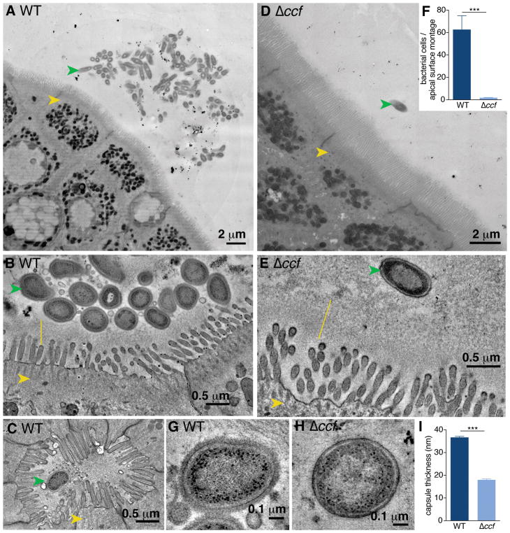
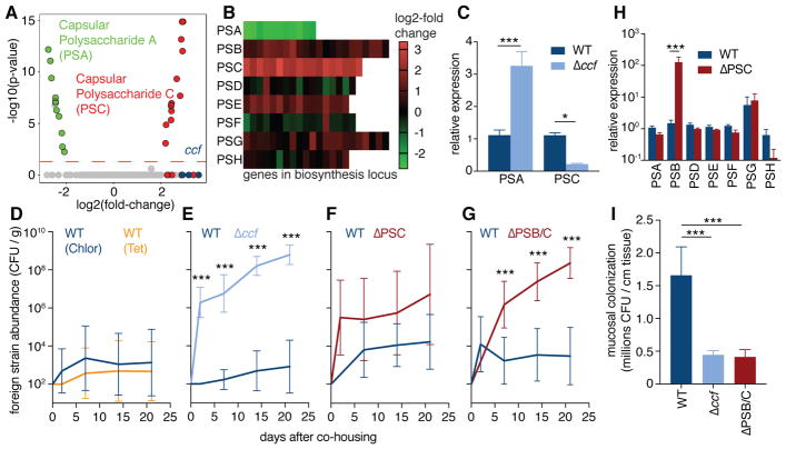
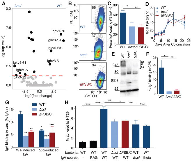
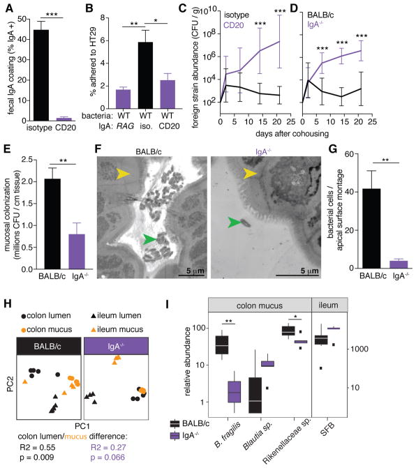
The immune system responds vigorously to microbial infection while permitting lifelong colonization by the microbiome. Mechanisms that facilitate the establishment and stability of the gut microbiota remain poorly described. We found that a regulatory system in the prominent human commensal Bacteroides fragilis modulates its surface architecture to invite binding of immunoglobulin A (IgA) in mice. Specific immune recognition facilitated bacterial adherence to cultured intestinal epithelial cells and intimate association with the gut mucosal surface in vivo. The IgA response was required for B. fragilis (and other commensal species) to occupy a defined mucosal niche that mediates stable colonization of the gut through exclusion of exogenous competitors. Therefore, in addition to its role in pathogen clearance, we propose that IgA responses can be co-opted by the microbiome to engender robust host-microbial symbiosis.


Figures

Fig. 1
Fig. 1. Bacteroides fragilis resides as aggregates on the colon epithelium in a CCF-dependent manner
(A) Representative transmission electron microscopy (TEM) projection and (B) high-resolution tomogram of epithelial-associated wild-type B. fragilis in mono-colonized mice. Ascending colons of mice harbored aggregates of B. fragilis (green arrow) under non-pathogenic conditions, that made tight associations with the glycocalyx (yellow line) overlying intestinal epithelial cells (IECs, yellow arrow). (C) Tomogram of wild-type B. fragilis penetrating deep into the duct of a crypt of Lieberkühn. (D) Representative TEM projection image and (E) tomogram of epithelial-associated B. fragilis Δccf. The absence of the CCF system abrogated formation of bacterial aggregates and prevented intimate association with the glycocalyx. (n = 3 mice per group, about 1 mm epithelium scanned per mouse). (F) Quantification of bacterial cells per projection montage (A and D) of epithelial-associated bacteria (unpaired t test, n = 7, 8 images from 4 mice per group). (G and H) Tomogram of the bacterial surface of wild-type B. fragilis (G) in comparison to B. fragilis Δccf (H) revealed a thick fuzzy capsule for wild-type bacteria residing in the colons of mice. (I) Measurement of capsule thickness (unpaired t test, n = 10 cells from 3 mice per group) (*** p < 0.001).
Fig. 2
Fig. 2. Specific capsular polysaccharides, regulated by ccf, are necessary for single-strain stability
(A) RNAseq gene expression analysis of B. fragilis overexpressing ccfA during laboratory culture growth, relative to empty vector control (n = 3). Green symbols represent PSA genes; red symbols represent PSC genes; blue symbols represent ccf genes. (B) Heat map of expression levels for all capsular polysaccharide loci in B. fragilis following ccfA overexpression during growth in culture. (C) Relative expression using qRT-PCR (ΔΔCt normalized to gyrase) of RNA from colon lumen contents of mice mono-colonized with B. fragilis or B. fragilis Δccf (Sidak 2-way ANOVA, n = 4). (D–G) Abundance of foreign strains exchanged between pairs of co-housed mice each mono-colonized with the indicated strains, in colony forming units (CFU) per gram of feces (Sidak repeated measure 2-way ANOVA on log-transformed data, geometric mean and 95% CI, n = 9–12 pairs per plot). (H) Relative expression levels of capsular polysaccharides analyzed by qRT-PCR (ΔΔCt normalized to gyrase) of RNA from colon lumen contents of mice mono-colonized with B. fragilis or B. fragilis ΔPSC (Sidak 2-way ANOVA, n = 3, 4). (I) Plating of CFU from ascending colon mucus of mice mono-colonized with B. fragilis strains (Tukey ANOVA, n = 8) (* p < 0.05, ** p < 0.01, *** p < 0.001).
Fig. 3
Fig. 3. B. fragilis induces a specific IgA response, dependent on ccf regulation of surface capsular polysaccharides, which enhances epithelial adherence
(A) RNAseq gene expression analysis of RNA recovered from whole ascending colon tissue of mice mono-colonized with B. fragilis or B. fragilis Δccf (n = 3). (B) Flow cytometry plots and (C) quantification of IgA coating of B. fragilis from feces of mice mono-colonized with various strains (Tukey ANOVA, n = 11–12). (D) ELISA for total fecal IgA in mono-colonized mice (Sidak repeated measure 2-way ANOVA, not significant, n = 4). (E) Bacterial lysates from feces of mono-colonized Rag1−/− mice probed in Western blots with fecal IgA from B. fragilis mono-colonized mice and (F) quantification of the proportional signal from IgA binding to capsular polysaccharides (CPS) (over 245 kDa) (Tukey ANOVA, n = 3 mice). (G) Binding of fecal IgA extracted from mono-colonized mice to various strains of B. fragilis. Source of IgA is mice colonized with either WT B. fragilis or B. fragilis Δccf. Because ccf is expressed in vivo, IgA-free bacteria from feces of mono-colonized Rag1−/− mice were used as the target for IgA binding (Tukey 2-way ANOVA, *significantly different from WT bacteria with WT IgA, n = 3). (H) In vitro epithelial cell adherence assay using IgA extracted from Swiss Webster mice (or Rag1−/−, second column) mono-colonized with B. fragilis or B. thetaiotaomicron (theta; last column). IgA-free but in vivo-adapted bacteria were isolated from mono-colonized Rag1−/− mice (Tukey ANOVA, n = 4 mice as the source of bacteria) (* p < 0.05, ** p < 0.01, *** p < 0.001).
Fig. 4
Fig. 4. IgA production in vivo is necessary for single-strain stability, mucosal colonization, and epithelial aggregation
(A) IgA coating of wild-type B. fragilis in feces following injection of anti-CD20 or isotype control antibody (unpaired t test, n = 8). (B) Epithelial cell adherence assay of wild-type B. fragilis incubated with IgA extracted from indicated mono-colonized mice (Tukey ANOVA, n = 4 mice as the source of bacteria). (C) Abundance of foreign strains exchanged between pairs of wild-type B. fragilis mono-colonized mice treated with anti-CD20 or an isotype control (Sidak repeated measure 2-way ANOVA on log-transformed data, n = 10). (D) Foreign strains exchanged between pairs of BALB/c and BALB/c IgA−/− mice mono-colonized with wild-type B. fragilis (Sidak repeated measure 2-way ANOVA on log-transformed data, n = 9). (E) CFU plating of ascending colon mucus of wild-type and IgA−/− mice mono-colonized with wild-type B. fragilis (unpaired t test, n = 9). (F) Representative TEM projections of ascending colon (yellow arrow: epithelial cell) from mice mono-colonized with wild-type B. fragilis (green arrow) (n = 3 mice per group, about 1 mm epithelium scanned per mouse) and (G) quantification of bacterial cells per projection montage (unpaired t test, n = 7, 6 images from 3 mice per group) (H) Principle coordinate analyses of weighed UniFrac distances of 16S community profiles of ex-germ-free BALB/c and BALB/c IgA−/− mice transplanted with a complex mouse microbiota (Adonis test within colon for lumen/mucus difference). (I) Relative abundance of B. fragilis and highly IgA-coated ESVs in ex-germ-free mice (* p < 0.05, ** p < 0.01, *** p < 0.001).

Comment in

  • Unlocking the secrets of IgA.
    Thomas H. Thomas H. Nat Rev Gastroenterol Hepatol. 2018 Jul;15(7):389. doi: 10.1038/s41575-018-0036-3. Nat Rev Gastroenterol Hepatol. 2018. PMID: 29849107 No abstract available.
  • Gut Microbiota: IgA Protects the Pioneers.
    Schofield WB, Palm NW. Schofield WB, et al. Curr Biol. 2018 Sep 24;28(18):R1117-R1119. doi: 10.1016/j.cub.2018.08.019. Curr Biol. 2018. PMID: 30253156

References

    1. Hall AB, Tolonen AC, Xavier RJ. Human genetic variation and the gut microbiome in disease. Nat Rev Genet. 2017 doi: 10.1038/nrg.2017.63. - DOI - PubMed
    1. Fung TC, Olson CA, Hsiao EY. Interactions between the microbiota, immune and nervous systems in health and disease. Nat Neurosci. 2017;20:145–155. - PMC - PubMed
    1. Faith JJ, et al. The long-term stability of the human gut microbiota. Science. 2013;341:1237439. - PMC - PubMed
    1. Truong DT, Tett A, Pasolli E, Huttenhower C, Segata N. Microbial strain-level population structure and genetic diversity from metagenomes. Genome Res. 2017 gr.216242.116. - PMC - PubMed
    1. Fagarasan S, et al. Critical roles of activation-induced cytidine deaminase in the homeostasis of gut flora. Science. 2002;298:1424–1427. - PubMed

Publication types