. 2018 May 1;9(5):513.
doi: 10.1038/s41419-018-0534-9.

HCC-derived exosomes elicit HCC progression and recurrence by epithelial-mesenchymal transition through MAPK/ERK signalling pathway

Affiliations

HCC-derived exosomes elicit HCC progression and recurrence by epithelial-mesenchymal transition through MAPK/ERK signalling pathway

Lu Chen et al. Cell Death Dis. .

Abstract

Liver cancer is the second most common cause of cancer-related death worldwide. Approximately 70-90% of primary liver cancers are hepatocellular carcinoma (HCC). Currently, HCC patient prognosis is unsatisfactory due to high metastasis and/or post-surgical recurrence rates. Therefore, new therapeutic methods for inhibiting metastasis and recurrence are urgently needed. Exosomes are small lipid-bilayer vesicles that are implicated in tumour development and metastasis. Rab27a, a small GTPase, regulates exosome secretion by mediating multivesicular endosome docking at the plasma membrane. However, whether Rab27a participates in HCC cell-derived exosome exocytosis is unclear. Epithelial-mesenchymal transition (EMT) frequently initiates metastasis. The role of HCC cell-derived exosomes in EMT remains unknown. We found that exosomes from highly metastatic MHCC97H cells could communicate with low metastatic HCC cells, increasing their migration, chemotaxis and invasion. Rab27a knockdown inhibited MHCC97H-derived exosome secretion, which consequently promoted migration, chemotaxis and invasion in parental MHCC97H cells. Mechanistic studies showed that the biological alterations in HCC cells treated with MHCC97H-derived exosomes or MHCC97H cells with reduced self-derived exosome secretion were caused by inducing EMT via MAPK/ERK signalling. Animal experiments indicated that exosome secretion blockade was associated with enhanced lung and intrahepatic metastasis of parental MHCC97H cells, while ectopic overexpression of Rab27a in MHCC97H cells could rescue this enhancement of metastasis in vivo. Injection of MHCC97H cell-derived exosomes through the tail vein promoted intrahepatic recurrence of HLE tumours in vivo. Clinically, Rab27a was positively associated with serum alpha-fetoprotein (AFP) level, vascular invasion and liver cirrhosis. Our study elucidated the role of exosomes in HCC metastasis and recurrence, suggesting that they are promising therapeutic and prognostic targets for HCC patients.


Conflict of interest statement

The authors declare that they have no conflict of interest.

Figures
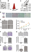
Fig. 1
Fig. 1. Highly metastatic MHCC97H-derived exosomes improve migration, chemotaxis and invasion of HLE cells.
a Transmission electron micrograph of MHCC97H exosomes. Arrows indicate exosomes and arrowheads indicate smaller nonexosomal vesicles. Scale bar, 200 nm. b The size distribution of isolated exosomes using Zetasizer Nano ZS90. c Equal amounts of total protein (60 μg) from MHCC97H cells and exosomes were analysed by Western blot for the presence of the exosomal markers CD63, Alix and TSG101, as well as AFP (a recognised HCC marker). The endoplasmatic reticulum protein GRP78 was used as the negative control. d Fluorescence microscopy images of HLE cells treated with fluorescently labelled MHCC97H exosomes (single-stranded RNAs: red; exosomal proteins: green) for 24 h. e Scratch assay for the migration of HLE cells treated with or without MHCC97H-derived exosomes (100 μg/ml). The distance was measured every 6 h for 24 h. f The chemotactic potential of HLE cells treated with or without MHCC97H-derived exosomes (100 μg/ml). The incubation time was 24 h. g Matrigel invasion assay for the invasion of HLE cells treated with or without MHCC97H-derived exosomes (100 μg/ml). The incubation time was 36 h. h Colony formation assay of HLE cells treated with or without MHCC97H-derived exosomes (100 μg/ml). The culture time was 2 weeks. Abbreviation: Exo exosome. *P < 0.05, **P < 0.01 and ***P < 0.001. Scale bar, 1.0 mm. Data are represented as the mean ± S.D. All experiments were repeated at least three times
Fig. 2
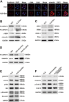
Fig. 2. Incubation of HLE cells with MHCC97H-derived exosomes promotes EMT through the MAPK/ERK pathway.
a Immunofluorescence microscopy of EMT markers in HLE cells treated with or without MHCC97H-derived exosomes for 24 h. b Western blot analysis of EMT markers in HLE cells treated with or without MHCC97H-derived exosomes for 24 h. c Western blot analysis of EMT promoters (ZEB1, ZEB2 and Slug) and MET promoter OVOL1 in HLE cells treated with or without MHCC97H-derived exosomes for 24 h. d Western blot analysis of EMT markers of HLE cells with exposure to MHCC97H-derived exosomes for different durations. e Western blot analysis of phosphorylated and total Erk1/2, Akt and Smad2/3 in HLE cells treated with or without MHCC97H-derived exosomes for 24 h. f Western blot analysis of EMT markers and phosphorylated and total Erk1/2 in HLE cells, exosome-treated HLE cells and exosome-treated HLE cells when cultured with the ERK inhibitor PD98059. Abbreviation: Exo exosome. All experiments were repeated at least three times
Fig. 3
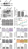
Fig. 3. Rab27a downregulation inhibits MHCC97H-derived exosome secretion and promotes migration, chemotaxis and invasion of parental cells.
a The knockdown efficiency of the Rab27a shRNA was examined by Western blot. b Equal amounts of total exosomal proteins (60 μg) secreted by siRab27a/MHCC97H and siSCR/MHCC97H cells were analysed by Western blot for the presence of the exosomal markers CD63, Alix and TSG101, as well as AFP (a recognised HCC marker). The endoplasmatic reticulum protein GRP78 was used as the negative control. GAPDH was used as loading control. c The total amounts of protein in exosome pellets purified from culture supernatant of MHCC97H (WT), siSCR/MHCC97H and siRab27a/MHCC97H cells were quantified by Bradford assay. d Alteration in migration by Rab27a knockdown was determined by scratch assays. e Alteration in chemotaxis by Rab27a knockdown was measured by chemotaxis assays. f Alteration in invasion by Rab27a knockdown was examined by Matrigel invasion assays. g Alteration in clonogenic ability by Rab27a knockdown was detected by colony formation assays. h Alteration in sphere formation ability by Rab27a knockdown was explored by 3D sphere formation assays. Abbreviation: Exo exosome. *P < 0.05, **P < 0.01 and ***P < 0.001. Scale bar, 1.0 mm. Data are represented as the mean ± S.D. All experiments were repeated at least three times
Fig. 4
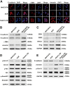
Fig. 4. Inhibition of self-derived exosome secretion by Rab27a blockade induces EMT through the MAPK/ERK pathway.
Analysis of the expression level of EMT markers in MHCC97H (WT), siSCR/MHCC97H and siRab27a/MHCC97H cells by immunofluorescence microscopy (a) and Western blot (b). c Analysis of the expression pattern of EMT-associated and MET-associated transcriptional regulators in MHCC97H (WT), siSCR/MHCC97H and siRab27a/MHCC97H cells at the protein level. d Western blot analysis of phosphorylated and total Erk1/2, Akt and Smad2/3 in MHCC97H (WT), siSCR/MHCC97H and siRab27a/MHCC97H cells. e Western blot analysis of phosphorylated and total Erk1/2 and EMT markers in MHCC97H (WT), siSCR/MHCC97H, and siRab27a/MHCC97H cells treated with or without the ERK inhibitor PD98059 for 24 h. All experiments were repeated at least three times
Fig. 5
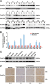
Fig. 5. Expression pattern of Rab27a in human HCCs and its correlation with clinicopathological features.
a Western blot analysis of Rab27a expression in 20 pairs of HCC tissues (C) and non-tumour tissues (N). b qRT-PCR of Rab27a expression in 11 pairs of HCC tissues and adjacent non-tumour tissues. c Western blot of the protein levels of Rab27a in 7 HCC cell lines and the human HL-7702 hepatocyte cell line. d Immunohistochemical staining analysis of Rab27a in non-tumour tissues and HCC tissues with varying degrees of vascular invasion. Three expression levels of staining: negative (−), weakly positive (+) and strongly positive (++); magnifications = ×20, ×40. Scale bar, 1.0 mm
Fig. 6
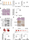
Fig. 6. Inhibition of self-derived exosome secretion or exogenous exosome uptake enhances HCC metastasis and recurrence in vivo.
a Comparison of tumour size and weight in nude mice subcutaneously implanted with siSCR/MHCC97H or siRab27a/MHCC97H cells (n = 4/group). b The lung metastatic ability of human tumours in the siSCR/MHCC97H cell-injection and siRab27a/MHCC97H cell-injection groups visualised using H&E staining, magnifications = ×20, ×40. The arrow indicates the metastatic foci. c Western blot analysis of Rab27a and EMT markers in subcutaneously formed tumours in the siSCR/MHCC97H cell-injection and siRab27a/MHCC97H cell-injection groups. d Immunohistochemical assay of E-cadherin and N-cadherin in subcutaneously formed tumours in the siSCR/MHCC97H cell-injection and siRab27a/MHCC97H cell-injection groups. e The intrahepatic metastatic ability of siRab27a/MHCC97H and siSCR/MHCC97H cells (n = 5). The thick arrow indicates the orthotopically implanted tumours, and the thin arrow indicated the intrahepatic metastatic foci. f The effect of MHCC97H-derived exosomes on intrahepatic recurrence of HLE tumours (n = 5). Abbreviation: Exo exosome. Data are represented as the mean ± S.D. *P < 0.05 and **P < 0.01. Scale bar, 1.0 mm
Fig. 7
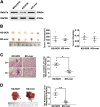
Fig. 7. Ectopic overexpression of Rab27a rescues HCC metastasis in vivo.
a The efficiency of Rab27a overexpression was examined by Western blot. b Comparison of tumour size and weight in nude mice subcutaneously implanted with overSCR-siRab27a/MHCC97H or overRab27a-siRab27a/MHCC97H cells (n = 5/group). c The lung metastatic ability of human tumours in the overSCR-siRab27a/MHCC97H cell-injection and overRab27a-siRab27a/MHCC97H cell-injection groups visualised using H&E staining, magnifications = ×20, ×40. The arrow indicates the metastatic foci. d The intrahepatic metastatic ability of overSCR-siRab27a/MHCC97H and overRab27a-siRab27a/MHCC97H cells (n = 5). The thick arrow indicates the orthotopically implanted tumours and the thin arrow indicated the intrahepatic metastatic foci. Abbreviation: KD-SCR overSCR-siRab27a/MHCC97H; KD-over overRab27a-siRab27a/MHCC97H. Data are represented as the mean ± S.D. *P < 0.05 and **P < 0.01. Scale bar, 1.0 mm

Similar articles

Cited by

References

    1. Torre LA, et al. Global cancer statistics, 2012. CA: Cancer J. Clin. 2015;65:87–108. - PubMed
    1. Wang FS, Fan JG, Zhang Z, Gao B, Wang HY. The global burden of liver disease: the major impact of China. Hepatology. 2014;60:2099–2108. doi: 10.1002/hep.27406. - DOI - PMC - PubMed
    1. Forner A, Llovet JM, Bruix J. Hepatocellular carcinoma. Lancet. 2012;379:1245–1255. doi: 10.1016/S0140-6736(11)61347-0. - DOI - PubMed
    1. Thery C, Zitvogel L, Amigorena S. Exosomes: composition, biogenesis and function. Nat. Rev. Immunol. 2002;2:569–579. doi: 10.1038/nri855. - DOI - PubMed
    1. Melo SA, et al. Cancer exosomes perform cell-independent microRNA biogenesis and promote tumorigenesis. Cancer Cell. 2014;26:707–721. doi: 10.1016/j.ccell.2014.09.005. - DOI - PMC - PubMed

Publication types

MeSH terms