. 2018 May 3;8(1):7009.
doi: 10.1038/s41598-018-24581-y.

Single vector platform vaccine protects against lethal respiratory challenge with Tier 1 select agents of anthrax, plague, and tularemia

Affiliations

Single vector platform vaccine protects against lethal respiratory challenge with Tier 1 select agents of anthrax, plague, and tularemia

Qingmei Jia et al. Sci Rep. .

Abstract

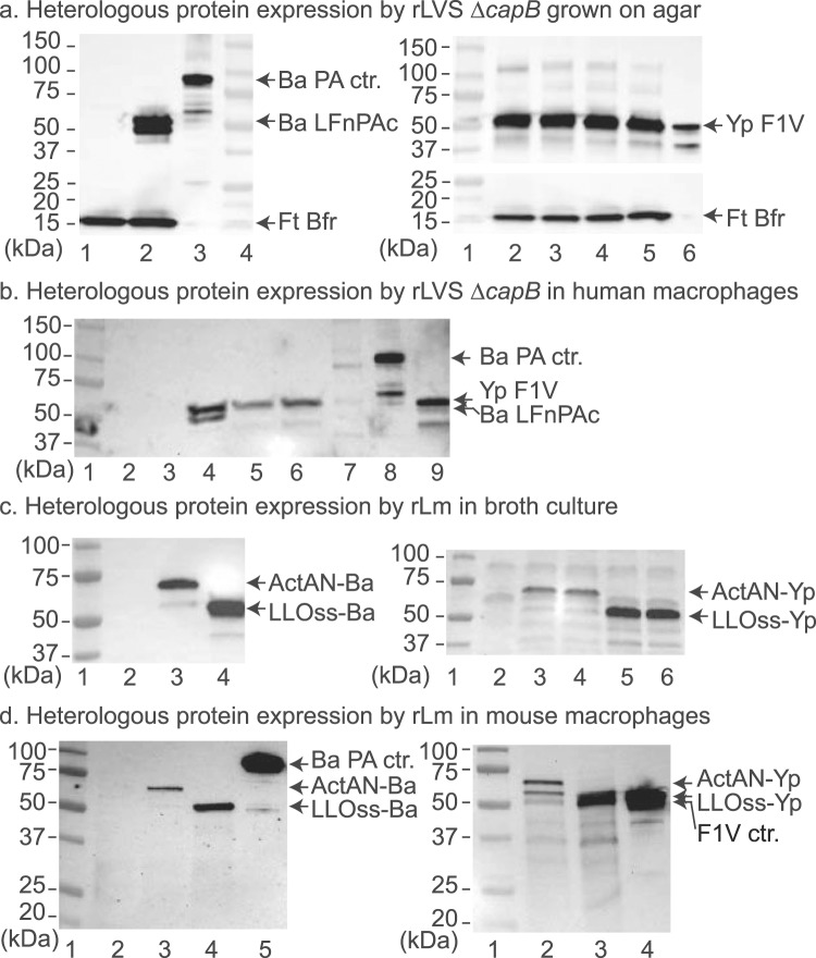
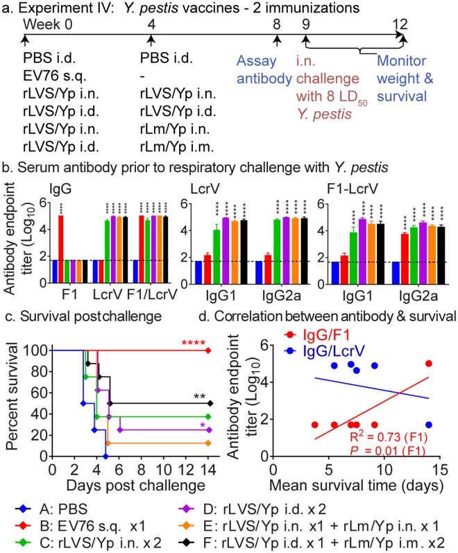
Bacillus anthracis, Yersinia pestis, and Francisella tularensis are the causative agents of Tier 1 Select Agents anthrax, plague, and tularemia, respectively. Currently, there are no licensed vaccines against plague and tularemia and the licensed anthrax vaccine is suboptimal. Here we report F. tularensis LVS ΔcapB (Live Vaccine Strain with a deletion in capB)- and attenuated multi-deletional Listeria monocytogenes (Lm)-vectored vaccines against all three aforementioned pathogens. We show that LVS ΔcapB- and Lm-vectored vaccines express recombinant B. anthracis, Y. pestis, and F. tularensis immunoprotective antigens in broth and in macrophage-like cells and are non-toxic in mice. Homologous priming-boosting with the LVS ΔcapB-vectored vaccines induces potent antigen-specific humoral and T-cell-mediated immune responses and potent protective immunity against lethal respiratory challenge with all three pathogens. Protection against anthrax was far superior to that obtained with the licensed AVA vaccine and protection against tularemia was comparable to or greater than that obtained with the toxic and unlicensed LVS vaccine. Heterologous priming-boosting with LVS ΔcapB- and Lm-vectored B. anthracis and Y. pestis vaccines also induced potent protective immunity against lethal respiratory challenge with B. anthracis and Y. pestis. The single vaccine platform, especially the LVS ΔcapB-vectored vaccine platform, can be extended readily to other pathogens.


Conflict of interest statement

This study was supported by National Institutes of Health grant AI101189. Flow cytometry was performed in the UCLA Jonsson Comprehensive Cancer Center (JCCC) and Center for AIDS Research Flow Cytometry Core Facility that is supported by National Institutes of Health awards P30 CA016042 and 5P30 AI028697, and by the JCCC, the UCLA AIDS Institute, and the David Geffen School of Medicine at UCLA. M.A.H. and Q.J. are inventors on patent applications filed by UCLA that include data presented herein.

Figures

Figure 1
Figure 1
Expression of heterologous fusion proteins of B. anthracis and Y. pestis by rLVS ΔcapB and rLm ΔactA ΔinlB prfA vaccines grown on agar and in infected macrophage-like cells. (a) Expression of B. anthracis (left) and Y. pestis (right) fusion proteins by rLVS ΔcapB grown on agar. Single colonies of chocolate agar grown rLVS ΔcapB/Ba and rLVS ΔcapB/Yp (4 clones) were lysed in SDS sample buffer and lysates analyzed by Western blotting using a mixture of antibody to B. anthracis PA and to F. tularensis Bfr (left panel) or antibody to Y. pestis LcrV protein followed by antibody to Bfr (right panel). Left panel, lane 1, LVS ΔcapB vector; lane 2, rLVS ΔcapB/Ba; lane 3, PA protein control; lane 4, protein mass standards. Right panel, lane 1, protein mass standards; lanes 2–5, rLVS ΔcapB/Yp; lane 6, monomer of F1-LcrV (F1V) protein control. (b) Expression of fusion proteins by rLVS ΔcapB in infected human macrophage-like cells. Monocytic THP-1 cells seeded on 24-well plates and differentiated in the presence of PMA were left uninfected or infected with LVS ΔcapB, rLVS ΔcapB/Ba or rLVS ΔcapB/Yp; cells were lysed at 24 h post infection, and cell lysates analyzed by Western blotting using a mixture of antibody to B. anthracis PA and to Y. pestis LcrV. Lanes 1 & 7, two different protein standards; lane 2, uninfected control; lane 3, LVS ΔcapB; lane 4, rLVS ΔcapB/Ba; lanes 5 and 6, two clones of rLVS ΔcapB/Yp vaccines; lane 8, B. anthracis PA and degraded proteins; lane 9, Y. pestis F1-LcrV monomer protein and degraded proteins. (c) Expression and secretion of heterologous fusion proteins by rLm vaccines in broth. Culture filtrates of Lm vector or rLm vaccines were analyzed by Western blotting using antibody to B. anthracis PA (left panel) or to Y. pestis LcrV (right panel). Left panel, lane 1, protein mass standards; lane 2, Lm vector; lane 3, rLm/ActAN-Ba; lane 4, rLm/LLOss-Ba. Right panel, lane 1, protein mass standards; lane 2, Lm vector; lanes 3 & 4, two clones of rLm/ActAN-Yp; lanes 5 & 6, two clones of rLm/LLOss-Yp. (d) Expression of heterologous fusion proteins by rLm vaccines in infected mouse macrophage-like cells. Monolayers of J774A.1 cells were not infected or infected with a stationary culture of rLm vaccines similarly as described above in the legend to b . Lysates were subjected to Western blotting analysis using antibody to B. anthracis PA (left) or to Y. pestis LcrV (right). Left panel, lane 1, protein standards; lane 2, uninfected control; lane 3, rLm/ActAN-Ba; lane 4, rLm/LLOss-Ba; lane 5, PA protein. Right panel, lane 1, protein standards; lane 2, rLm/ActAN-Yp; lane 3, rLm/LLOss-Yp; lane 4, F1-LcrV protein control. (ad) On the left border of each panel are listed the masses of protein standards; on the right border are listed the proteins of interest. Each blot was processed by using the Bio-Rad imaging system (ChemiDoc XRS) and Quantity One software, which allows the overlap of a white-light image, for visualization of the protein standards (a, left panel lane 4 and right panel lane 1; bd, lane 1), and a chemiluminescent image, for visualization of the antibody-labeled protein bands. The full-length blots in panels b–d are shown in the Supplementary Information (Fig. S1).
Figure 2
Figure 2
Two immunizations by homologous priming-boosting with rLVS ΔcapB/Ba or by heterologous priming-boosting with rLVS ΔcapB/Ba - rLm ΔactA ΔinlB prfA/Ba induce humoral immune responses and protective immunity against respiratory challenge with virulent B. anthracis Ames spores. (a) Experiment schedule. Mice (n = 8/group) were immunized homologously twice, 4 weeks apart, with PBS intranasally (i.n.) (Sham), AVA (0.025 ml) subcutaneously (s.q.), or 106 CFU rLVS ΔcapB/Ba (rLVS/Ba) i.n. or intradermally (i.d.), or heterologously with 106 CFU rLVS ΔcapB/Ba i.n. or i.d. and rLm/Ba i.n. or intramuscularly (i.m.), 4 weeks apart; bled at Week 7; challenged at week 8 with B. anthracis Ames spores (205,000/mouse, ~5 LD50); and monitored for survival for three weeks, as indicated. (b) Serum antibody after vaccination. Sera were assayed for antibody endpoint titer to B. anthracis PA and LF antigens and to F. tularensis heat-inactivated LVS (HI-LVS). Values are mean + SEM of serum antibody endpoint titer for n = 8 mice per group. Differences among individual groups were evaluated by Two-way ANOVA with Tukey’s correction. Values significantly different from the Sham group are marked with asterisks over the comparison groups. *P < 0.05; **P < 0.01; ***P < 0.001; ****P < 0.0001. (c) Survival after vaccination and challenge. The survival curve of each vaccinated group is compared with that of Group A (Sham) or Group B (AVA) by the log-rank test (Mantel-cox); P values for individual vaccine groups significantly different from the Sham or AVA group are marked with asterisks and “§”, respectively, color-coded to the color of the vaccine symbol; **P < 0.01; ***P < 0.001 vs. Sham group; §P < 0.05 vs. AVA group. (d) Correlation between serum antibody and mean survival time. Linear regression was used to obtain values for the slope and intercept and the correlation coefficient (R2) between pre-challenge serum antibody and post-challenge mean survival time at 21 days post-challenge. Two-tailed P values were calculated for the correlation.
Figure 3
Figure 3
Three immunizations by homologous priming-boosting with rLVS ΔcapB/Ba or by heterologous priming-boosting with rLVS ΔcapB/Ba – rLm ΔactA ΔinlB prfA/Ba induce high-level antibody responses and potent protective immunity against virulent B. anthracis respiratory challenge. (a) Experiment schedule. Mice (n = 8/group) were immunized two or three times homologously with 106 CFU rLVS ΔcapB/Ba (rLVS/Ba) or rLm ΔactA ΔinlB prfA/Ba (rLm/Ba) or heterologously first with rLVS/Ba and subsequently with rLm/Ba, as indicated. Controls were sham-immunized with PBS i.d. or with AVA s.q. three times. All mice were bled at week 11; challenged at week 12 with B. anthracis Ames spores (371,000 CFU, ~10 LD50); and monitored for survival for 3 weeks post-challenge. (b) Serum antibody prior to challenge and survival post challenge. Top panels. Sera were assayed for IgG or IgG subtypes IgG1 and IgG2a to B. anthracis PA and LF proteins, as indicated, after homologous (left two panels) and heterologous (right two panels) prime-boost vaccination. Values are mean + SEM of serum antibody endpoint titer for n = 8 per group. Differences in serum endpoint titer among individual groups were analyzed by two-way ANOVA with Tukey’s corrections. *P < 0.05; **P < 0.01; ****P < 0.0001 vs. Sham group. Bottom panels. The survival curve of each vaccinated group, color-coded as indicated in panel b, after homologous (left) and heterologous (right) prime-boost vaccination and challenge is compared with that of the Sham group by the log-rank test (Mantel-cox); P values for vaccine groups that are significantly different from the Sham group are marked with one or more asterisks color-coded to the color of the vaccine symbol. *P < 0.05 and **P < 0.01. (c) Correlation between serum antibody and mean survival time. The correlation coefficient (R2) and one-tailed P values were obtained as described in legend to Fig. 2d.
Figure 4
Figure 4
Homologous prime-boost vaccination with rLVS ΔcapB/Ba or heterologous prime-boost vaccination with rLVS ΔcapB/Ba - rLm ΔactA ΔinlB prfA/Ba induces antigen-specific cell-mediated immune responses. (a) Experiment schedule. Mice (n = 4/group) were immunized homologously with PBS i.n. (Sham), AVA s.q., or 106 CFU rLVS ΔcapB/Ba (rLVS/Ba) i.n., or heterologously with rLVS/Ba i.n. followed by 106 CFU rLm ΔactA ΔinlB prfA/Ba (rLm/Ba) i.n. or i.m. as indicated. At week 7, all mice were bled, euthanized, and their lung and spleen cells assayed for cytokine secretion and intracellular cytokine staining. (b) Antigen-specific cytokine secretion. Single cell suspensions of lung and spleen cells were stimulated with B. anthracis PA or LF proteins or F. tularensis heat-inactivated LVS (HI-LVS) for 3 days, as indicated, and cell supernatants assayed for interferon gamma (IFN-γ) (left two panels) and IL-4 (right two panels) by ELISA. Shown are the amounts of IFN-γ and IL-4 in the culture supernate in response to PA, LF, and HI-LVS. (c,d) Cytokine-expressing CD4+ (c) and CD8+ (d) T cells. Lung and spleen cells were stimulated with LF, PA, or HI-LVS as indicated at the top of the panels (c) or on the horizontal axis (d) and assayed by intracellular cytokine staining for CD4+ (c) and CD8+ (d) T cells expressing IFN-γ, TNF-α, IL-2, and/or IL-17A. Shown are the frequencies of CD4+ T cells expressing IFN-γ, TNF-α, IL-2, and/or IL-17A (c) and CD8+ T cells expressing IFN-γ (d). Values in bd are means + SEM. Differences among individual groups were evaluated by Two-way ANOVA with Tukey’s correction. Values significantly different from the Sham group are marked with asterisk(s) over brackets above the comparison groups; *P < 0.05; **P < 0.01; ***P < 0.001; ****P < 0.0001. Results shown are representative of three similar experiments.
Figure 5
Figure 5
Heterologous prime-boost vaccination with rLVS ΔcapB/Ba - rLm ΔactA ΔinlB prfA/Ba induces elevated frequencies of LF- and PA-specific polyfunctional lung and spleen CD4+ T-cells producing IFN-γ, IL-2, TNF-α, and IL-17. As described in Fig. 4a and indicated by the colors and letters at the bottom of the figure, mice (n = 4/group) were sham-immunized or immunized with various vaccines. Lung (left panels) and spleen (right panels) cells were stimulated with LF (a,b), PA (c,d), HI-LVS (e,f), or PMA (g,h) and assayed by intracellular cytokine staining for 15 possible combinations of CD4+ T-cells expressing IFN-γ, TNF-α, IL-2, and/or IL-17A. Values are means + SEM and differences among individual groups were analyzed by Two-way ANOVA with Tukey’s multiple comparisons test (Prism). Values significantly different from the Sham group are marked with asterisks over the comparison groups. In panels e, f and g, the Sham group is indicated by a short bar with an open end and the comparison groups are indicated by a short vertical line across the short bar. *P < 0.05; **P < 0.01; ***P < 0.001; ****P < 0.0001. Results shown are representative of three similar experiments.
Figure 6
Figure 6
Two immunizations by homologous priming-boosting with rLVS ΔcapB/Yp or heterologous priming-boosting with rLVS ΔcapB - rLm ΔactA ΔinlB prfA/Yp induces humoral immune responses and protective immunity against respiratory challenge with virulent Y. pestis. (a) Experiment schedule. Mice (n = 8/group) were immunized homologously twice with PBS (Sham) or 106 CFU LVS ΔcapB/Yp (rLVS/Yp) or once with 106 CFU EV76, or heterologously first with 106 CFU rLVS ΔcapB/Yp and then with 106 CFU rLm ΔactA ΔinlB prfA/Yp (rLm/Yp). All mice were bled at Week 8; challenged at week 9 with virulent Y. pestis (CO92) (1,900 CFU/mouse, ~8 LD50); and monitored for survival for three weeks, as indicated. (b) Serum antibody after vaccination. Sera were assayed for IgG antibody specific to F1, LcrV, or the monomer of F1-LcrV proteins (leftmost panel) and subtypes IgG1 and IgG2a antibodies specific to LcrV or F1-LcrV (middle and rightmost panels, resp.). Data are mean + SEM of serum antibody endpoint titer for n = 8 per group. Differences among individual groups were analyzed by two-way ANOVA with Tukey’s correction. ****P < 0.0001 vs. Sham. (c) Survival after challenge. The survival curve of each vaccinated group is compared with the Sham group (Group A) by the log-rank test (Mantel-cox); P values that are significantly different from the Sham group are marked with one or more asterisks color-coded to the color of the vaccine symbol. There were no changes in percent survival of mice in any group after day 14 post challenge until the end of the experiment. *P < 0.05; **P < 0.01; ***P < 0.001; ****P < 0.0001. (d) Correlation between serum antibody and mean survival time. The correlation coefficient (R2) and one-tailed P values were obtained as described in legend to Fig. 2D.
Figure 7
Figure 7
Three systemic immunizations by homologous priming-boosting with LVS ΔcapB/Yp induce high level antibody responses and potent protective immunity against respiratory challenge with virulent Y. pestis CO92 strain. (a) Experiment schedule. Mice (n = 8/group) were immunized homologously three times with 106 CFU rLVS ΔcapB/Yp (rLVS/Yp) or with 106 CFU rLm ΔactA ΔinlB prfA/Yp (rLm/Yp) or heterologously with rLVS ΔcapB/Yp and subsequently rLm/Yp. Mice vaccinated with PBS i.d. (Sham, Group A) or 106 CFU Y. pestis vaccine strain EV76 served as controls. All the mice were bled at Week 11; challenged with Y. pestis CO92 (1,800 CFU/mouse, ~7 LD50) at Week 12; and monitored for survival for 3 weeks post-challenge, as indicated. (b) Serum antibody after vaccination. Sera were assayed for antibody endpoint titer of IgG or IgG subtypes IgG1 and IgG2a to Y. pestis F1 (left panel) and LcrV (right panel) proteins. Data are mean + SEM of serum antibody endpoint titer for n = 8 per group. Differences among individual groups were analyzed by two-way ANOVA with Tukey’s correction. ***P < 0.001; ****P < 0.0001 vs. Sham. (c) Survival after vaccination and challenge. The survival curve of each vaccinated group is compared with that of the Sham group by the log-rank test (Mantel-cox); P values for vaccine groups significantly different from the Sham group are marked with one or more asterisks color-coded to the color of the vaccine symbol. *P < 0.05; ***P < 0.001. (d) Correlation between serum antibody and mean survival time. The correlation coefficient (R2) and one-tailed P values were obtained as described in legend to Fig. 2D.
Figure 8
Figure 8
Two immunizations by homologous priming-boosting with rLVS ΔcapB/iglABC induces strong protection against respiratory challenge with the virulent F. tularensis Schu S4 strain. (a) Schedule - Experiment VI. Mice (n = 8/group) were immunized once with PBS (Sham), 104 CFU LVS, 106 CFU LVS ΔcapB vector, or 106 CFU rLVS ΔcapB/iglABC; or twice with 106 CFU rLVS ΔcapB/iglABC; challenged i.n. with F. tularensis Schu S4 (10 CFU, ~10 LD50) at Week 10; and monitored for signs of illness, weight change, and death for 3 weeks, as indicated. (b) Survival after vaccination and challenge – Experiment VI. The survival curve of each vaccinated group is compared with that of the Sham (Group A) or LVS ΔcapB vector (Group C) group by the log-rank test (Mantel-cox); P values for vaccine groups that are significantly different from the Sham or LVS ΔcapB vector control group are marked with asterisk(s) and multiple “§”, respectively, color-coded to the color of the vaccine symbol. *P < 0.05; ***P < 0.001; and ****P < 0.0001 vs. Group A (Sham); §§P < 0.01; §§§P < 0.001 vs. Group C (vector control). (c) Schedule - Experiment VII. Mice (n = 8/group) were immunized once with PBS (Sham), 104 CFU LVS, or 106 CFU rLVS ΔcapB/iglABC three times at Weeks 0, 4, and 8, or twice at Weeks 4 and 8, bled at Week 13, and challenged i.n. with 2 CFU (2 LD50) or 6 CFU (6 LD50) F. tularensis Schu S4 at Week 14, and monitored for 3 weeks, as indicated. (d) Survival after vaccination and challenge and serum antibody pre-challenge – Experiment VII. Upper panels – Survival. The survival curve after challenge with 2 LD50 (left panel) or 6 LD60 (right panel) of each vaccinated group is compared with that of either the sham- or LVS-immunized group by the log-rank test (Mantel-cox); P values that are significantly different from the control group are marked with asterisk(s) color-coded to the color of the vaccine symbol. ***P < 0.001 vs. Group A (Sham); *P < 0.05 vs. Group B (LVS). Lower panel- Serum antibody after vaccination. Sera were assayed for IgG and subtypes IgG1 and IgG2a specific to heat-inactivated LVS. Data are mean + SEM of serum antibody endpoint titer for n = 8 per group. Differences among individual groups were compared by two-way ANOVA with Tukey’s correction. Values that are significantly different between two groups are marked with asterisk(s) over an open horizontal line crossing above the two groups. As indicated by the asterisks above the Sham group, its titers were significantly different from all other groups. *P < 0.05; **P < 0.01; and ****P < 0.0001.

Similar articles

Cited by

References

    1. Jernigan DB, et al. Investigation of bioterrorism-related anthrax, United States, 2001: epidemiologic findings. Emerg Infect Dis. 2002;8:1019–1028. doi: 10.3201/eid0810.020353. - DOI - PMC - PubMed
    1. Rosenzweig JA, et al. Progress on plague vaccine development. Appl Microbiol Biotechnol. 2011;91:265–286. doi: 10.1007/s00253-011-3380-6. - DOI - PubMed
    1. Matyas BT, Nieder HS, Telford SR., III. Pneumonic tularemia on Martha’s Vineyard: clinical, epidemiologic, and ecological characteristics. Ann N Y Acad Sci. 2007;1105:351–377. doi: 10.1196/annals.1409.013. - DOI - PubMed
    1. Alibek K. Biohazard: the chilling true story of the largest covert biological weapons program in the world, told from the inside by the man who ran it. Random House, Inc. (1999).
    1. Inglesby TV, et al. Plague as a biological weapon: medical and public health management. Working Group on Civilian Biodefense. JAMA. 2000;283:2281–2290. doi: 10.1001/jama.283.17.2281. - DOI - PubMed

Publication types

MeSH terms