. 2018 Apr 19:11:123.
doi: 10.3389/fnmol.2018.00123. eCollection 2018.

Fine Localization of Acetylcholinesterase in the Synaptic Cleft of the Vertebrate Neuromuscular Junction

Affiliations

Fine Localization of Acetylcholinesterase in the Synaptic Cleft of the Vertebrate Neuromuscular Junction

Edna Blotnick-Rubin et al. Front Mol Neurosci. .

Abstract

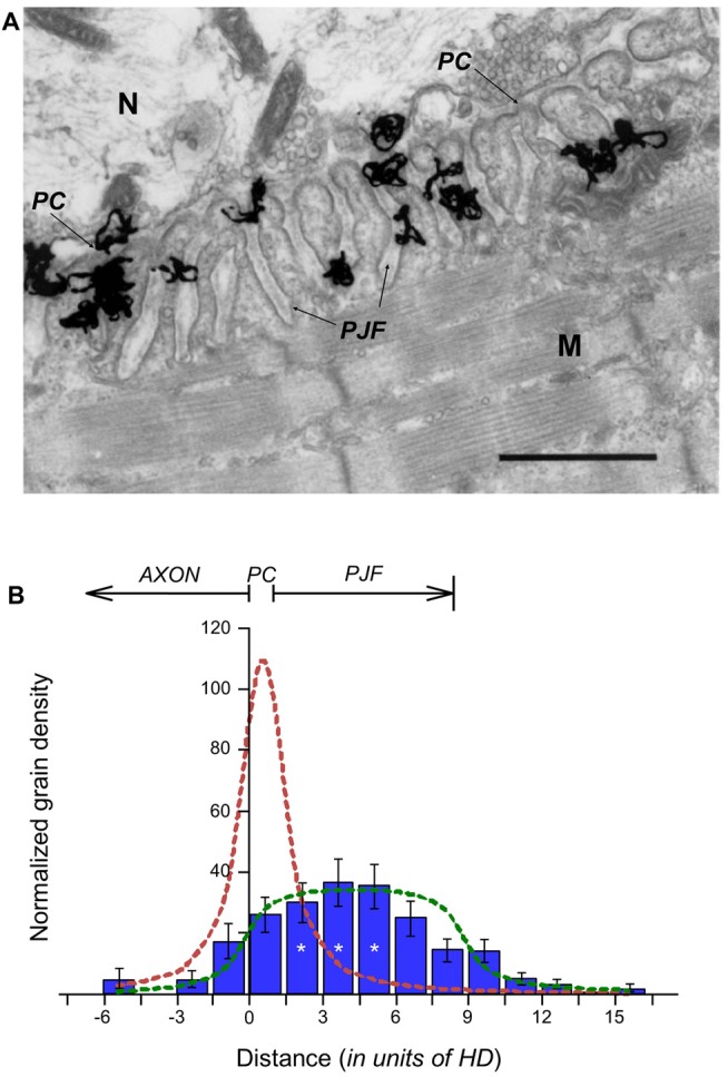
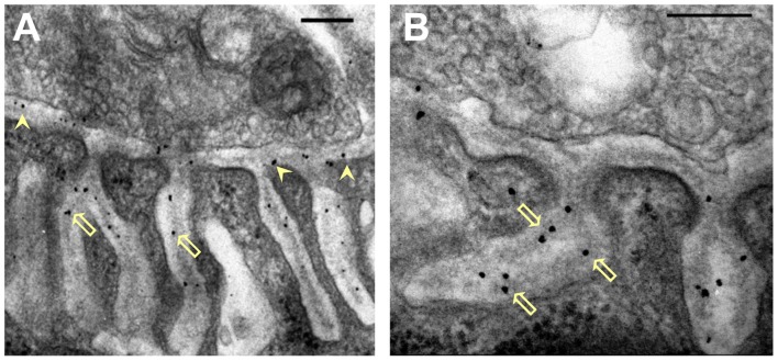
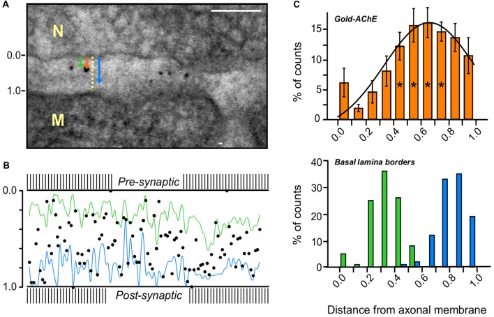
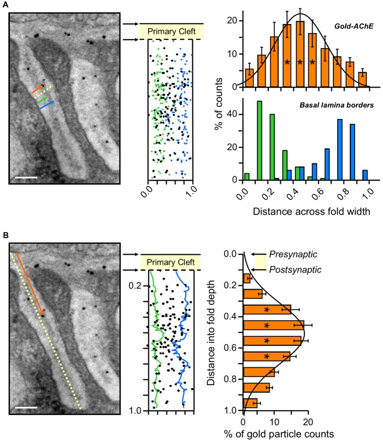
Acetylcholinesterase (AChE) is concentrated at cholinergic synapses, where it is a major factor in controlling the duration of transmitter action. The concentration and localization of AChE within the synaptic cleft are in keeping with the functional requirements of the particular type of synapse. The densities of synaptic AChE at various neuromuscular junctions (NMJs) had been evaluated by quantitative EM-autoradiography using radiolabeled probes. Yet, fundamental issues concerning the precise distribution and location of the enzyme in the cleft remained open: whether and to what extent synaptic AChE is associated with pre- or postsynaptic membranes, or with synaptic basal lamina (BL), and whether it occurs only in the primary cleft (PC) or also in postjunctional folds (PJFs). Nanogold-conjugates of fasciculin, an anticholinesterase polypeptide toxin, were prepared and used to label AChE at NMJs of mouse and frog muscles. Selective intense labeling was obtained at the NMJs, with gold-labeled AChE sites distributed over the BL in the PC and the PJFs. Quantitative analysis demonstrated that AChE sites are almost exclusively located on the BL rather than on pre- or postsynaptic membranes and are distributed in the PC and down the PJFs, with a defined pattern. This localization pattern of AChE is suggested to ensure full hydrolysis of acetylcholine (ACh) bouncing off receptors, thus eliminating its unnecessary detrimental reattachment.

Keywords: acetylcholinesterase; basal lamina; nanogold; postjunctional folds; synaptic cleft.


Figures

Figure 1
Figure 1
Distribution of Acetylcholinesterase (AChE) sites within the synaptic cleft. (A) EM-autoradiogram of mouse neuromuscular junction (NMJ) labeled with 125I-Fas. Mouse sternomastoid muscles were incubated with 125I-Fas (0.4 mM, 55 Ci/mmole) to inhibit AChE activity. They were then rinsed, fixed and processed for EM-autoradiography. Grains appeared almost exclusively at the NMJs. N, nerve; M, muscle. Arrows point to the primary cleft (PC) and to postsynaptic junctional folds (PJFs). Bar, 1 μm. (B) Radioactivity is distributed over the full depth of the PJFs. Grain density histogram (grains per unit area of autoradiogram) as a function of distance (in resolution units of HD = 80 nm; see, Salpeter et al., 1977) on either side of the axonal membrane (at 0), of 81 NMJs, three mice: left side into axon; right side into muscle. The bottom of the PJFs is on the average 11 HD (arrow), but with a large range; consequently, it is not as sharp a boundary as that provided by the axonal membrane. The reduced grain densities on both sides of the histogram are consistent with the radiation sources being within the PC and PJF, the variability in the depths of the PJFs and the actual spread of radiation under the experimental conditions (>3 HD). *, see text in “Results” section for detailed statistical parameter.
Figure 2
Figure 2
Distribution of nanogold-labeled AChE sites in the synaptic cleft of the mouse endplate. (A,B) Electron micrographs of cross sections through endplates of mouse sternomastoid muscles that had been incubated with Fas-biotin followed by 1.4 nm-nanogold-streptavidin and light silver intensification (details under “Materials and Methods” section). Gold particles (black) are seen over the PC (A, arrowheads) and PJFs, and are predominantly associated with the basal lamina (BL; B, hollow arrows). Bars, 0.2 μm.
Figure 3
Figure 3
AChE distribution in the PC of a mouse sternomastoid endplate. (A) Paradigm for the measurement of the distribution of gold-labeled AChE sites in the PC. Distances from the axonal pre-synaptic membrane (0.0) of each gold particle (orange arrow), and of the boundaries of the BL on both sides of the particle (green and blue arrows), were measured and divided by the width of the PC at that site (yellow dashed line); Bar, 0.1 μm. (B) Cumulative display of the gold-labeled AChE sites (measured in A) in a normalized PC of mouse sternomastoid endplate (five mice, 28 NMJs). Most gold particles are found on the synaptic BL (blue and green lines connect the BL border measurements on either side) in the PC between the presynaptic axonal membrane (0.0) and the postsynaptic muscle membrane (1.0). (C) Quantitative distribution of the gold-labeled AChE sites across the width of the PC, from compiled data obtained as in (A,B). Presynaptic axonal membrane, at X = 0.0, postsynaptic muscle membrane at X = 1.0. The histogram of the gold-particles (orange, upper panel) relative to the histograms of the BL boundaries (green-blue, lower panel) shows that the AChE sites are mainly spread over the BL in the PC. It should be noted that the AChE sites are predominantly closer to the postsynaptic muscle membrane than to the presynaptic nerve membrane. *, see text in “Results” section for detailed statistical parameters.
Figure 4
Figure 4
Gold-AChE distribution in the PJF. (A) Distribution across the width of the PJF. Left panel, measurement paradigm: distances from the myofiber fold membrane of each gold particle (orange arrow) and of the BL boundaries (green and blue arrows) were measured and divided by the width of the fold at that site (yellow dashed line). Center panel, compiled locations of the gold-labeled AChE sites (black) positioned in a normalized cleft within the adjacent measured BL boundaries (green and blue, as in A). Right panel, histograms of the compiled gold-labeled AChE sites (top, orange) relative to the adjacent BL boundaries (bottom, green and blue) reveal a spread of AChE sites over the BL within the PJF. Gaussian fit shows maximal concentration of sites at the center of the BL, with an even distribution towards either side. Fold width, X = 0.0 to X = 1.0. (B) Distribution along the long axis of the PJF. Left panel, measurement paradigm: The distance of each gold particle from the axonal membrane (orange arrow) was divided by the total depth of the fold (yellow dashed line). Center panel, compiled locations of gold-labeled AChE sites (black) in a normalized cleft within the outlined green and blue BL boundaries, marked above (A, center), and smoothed by moving average for convenient visualization. Right panel, histogram of the compiled data for the distances of the gold-labeled AChE sites from the axonal membrane (left panel) shows the distribution of AChE sites along the length of the PJF, with a maximum about halfway down. Data accumulated from micrographs of same NMJs as in Figure 3 (five mice, 28 NMJs). Axonal membrane, X = 0.0; Postsynaptic muscle membrane (top of fold), X = 0.1; Bottom of fold, X = 1.0. *, see text in “Results” section for detailed statistical parameters. Bars, 0.1 μm.
Figure 5
Figure 5
Distribution of gold-labeled AChE sites in the synaptic cleft of the frog NMJ. Frog cutaneous pectoris muscles were incubated with nanogold (1.4 nm) conjugated directly with Fas to label AChE (A,B), or with biotin-α-bungarotoxin followed by nanogold-streptavidin to label nAChR (C). (A) Nanogold-Fas conjugate labeled exclusively the NMJs, within both the PC and the PJFs. (B) An enlarged synaptic area (framed in A) shows the association of the gold-labeled AChE sites with the BL in both the PC and the PJFs. (C) In contrast, labeling of nAChR sites with the nanogold-α-bungarotoxin conjugate is seen to be at the crests and mouths of the PJFs. N, nerve; M, muscle; Arrowheads, labeled AChE in PC; Hollow arrows, labeled AChE in PJF; White arrowhead in (C), labeled nAChR (on postsynaptic membrane). Bars, 0.2 μm.

References

    1. Anglister L. (1991). Acetylcholinesterase from the motor nerve terminal accumulates on the synaptic basal lamina of the myofiber. J. Cell Biol. 115, 755–764. 10.1083/jcb.115.3.755 - DOI - PMC - PubMed
    1. Anglister L., McMahan U. J. (1985). Basal lamina directs acetylcholinesterase accumulation at synaptic sites in regenerating muscle. J. Cell Biol. 101, 735–743. 10.1083/jcb.101.3.735 - DOI - PMC - PubMed
    1. Anglister L., Eichler J., Szabo M., Haesaert B., Salpeter M. M. (1998). 125I-labeled fasciculin 2: a new tool for quantitation of acetylcholinesterase densities at synaptic sites by EM-autoradiography. J. Neurosci. Methods 81, 63–71. 10.1016/s0165-0270(98)00015-6 - DOI - PubMed
    1. Anglister L., Haesaert B., McMahan U. J. (1994a). Globular and asymmetric acetylcholinesterase in the synaptic basal lamina of skeletal muscle. J. Cell Biol. 125, 183–196. 10.1083/jcb.125.1.183 - DOI - PMC - PubMed
    1. Anglister L., Stiles J. R., Salpeter M. M. (1994b). Acetylcholinesterase density and turnover number at frog neuromuscular junctions, with modeling of their role in synaptic function. Neuron 12, 783–794. 10.1016/0896-6273(94)90331-x - DOI - PubMed

LinkOut - more resources