. 2018 May;15(5):6562-6570.
doi: 10.3892/ol.2018.8154. Epub 2018 Mar 2.

Exogenous hydrogen sulfide promotes hepatocellular carcinoma cell growth by activating the STAT3-COX-2 signaling pathway

Affiliations

Exogenous hydrogen sulfide promotes hepatocellular carcinoma cell growth by activating the STAT3-COX-2 signaling pathway

Yulan Zhen et al. Oncol Lett. 2018 May.

Abstract

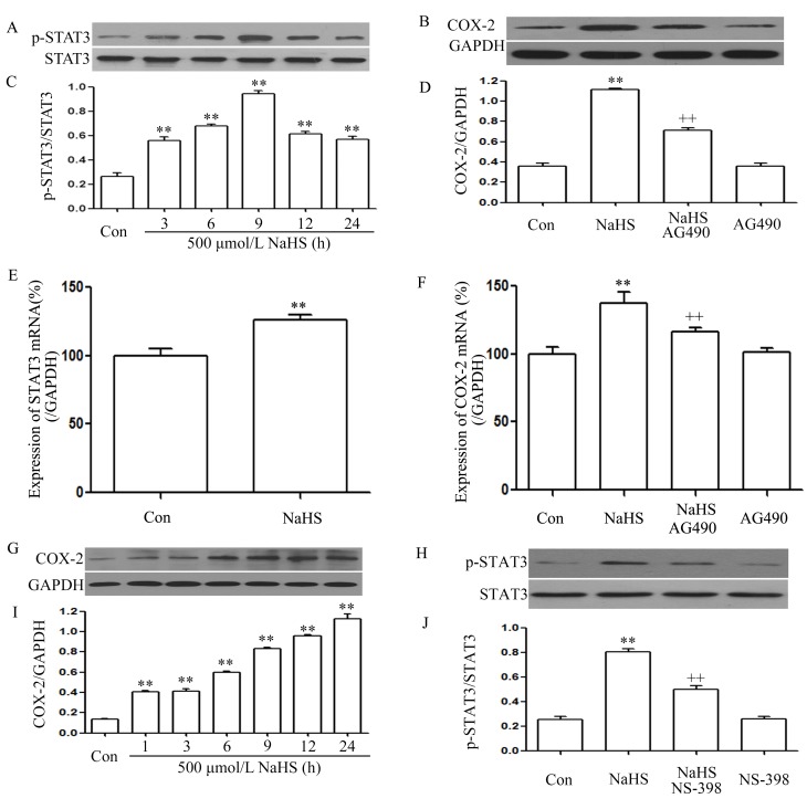
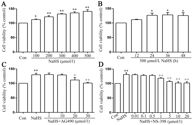
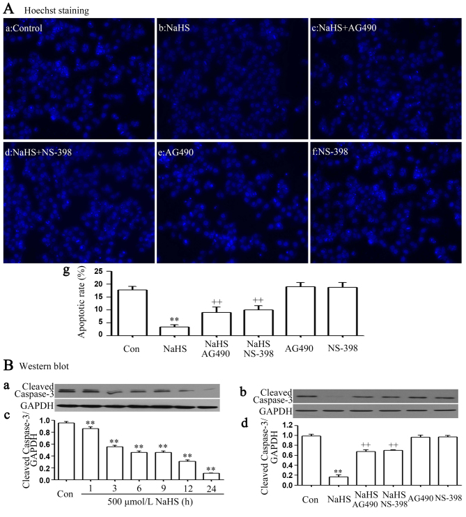
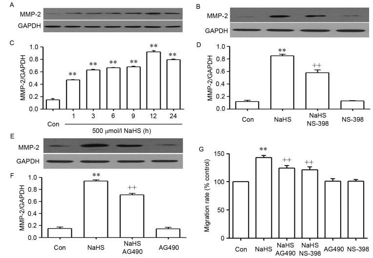
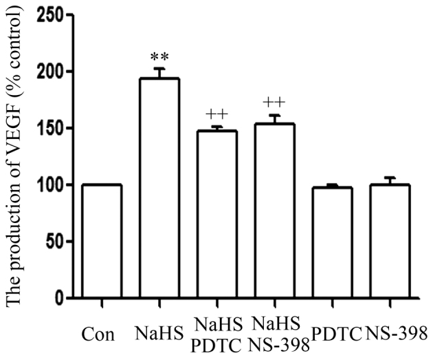
The effects of hydrogen sulfide (H2S) on cancer are controversial. Our group previously demonstrated that exogenous H2S promotes the development of cancer via amplifying the activation of the nuclear factor-κB signaling pathway in hepatocellular carcinoma (HCC) cells (PLC/PRF/5). The present study aimed to further investigate the hypothesis that exogenous H2S promotes PLC/PRF/5 cell proliferation and migration, and inhibits apoptosis by activating the signal transducer and activator of transcription 3 (STAT3)-cyclooxygenase-2 (COX-2) signaling pathway. PLC/PRF/5 cells were treated with 500 µmol/l NaHS (a donor of H2S) for 24 h. The expression levels of phosphorylated (p)-STAT3, STAT3, cleaved caspase-3 and COX-2 were measured by western blot assay. Cell viability was detected by Cell Counting kit-8 assay. Apoptotic cells were observed by Hoechst 33258 staining. The expression of STAT3 and COX-2 messenger RNA (mRNA) was detected by semiquantitative reverse transcription-polymerase chain reaction. The production of vascular endothelial growth factor (VEGF) was evaluated by ELISA. The results indicated that treatment of PLC/PRF/5 cells with 500 µmol/l NaHS for 24 h markedly increased the expression levels of p-STAT3 and STAT3 mRNA, leading to COX-2 and COX-2 mRNA overexpression, VEGF induction, decreased cleaved caspase-3 production, increased cell viability and migration, and decreased number of apoptotic cells. However, co-treatment of PLC/PRF/5 cells with 500 µmol/l NaHS and 30 µmol/l AG490 (an inhibitor of STAT3) or 20 µmol/l NS-398 (an inhibitor of COX-2) for 24 h significantly reverted the effects induced by NaHS. Furthermore, co-treatment of PLC/PRF/5 cells with 500 µmol/l NaHS and 30 µmol/l AG490 markedly decreased the NaHS-induced increase in the expression level of COX-2. By contrast, co-treatment of PLC/PRF/5 cells with 500 µmol/l NaHS and 20 µmol/l NS-398 inhibited the NaHS-induced increase in the expression level of p-STAT3. In conclusion, the findings of the present study provide evidence that the STAT3-COX-2 signaling pathway is involved in NaHS-induced cell proliferation, migration, angiogenesis and anti-apoptosis in PLC/PRF/5 cells, and suggest that the positive feedback between STAT3 and COX-2 may serve a crucial role in hepatocellular carcinoma carcinogenesis.

Keywords: COX-2; PLC/PRF/5 cells; STAT3; growth; hydrogen sulfide.


Figures

Figure 1.
Figure 1.
NaHS activates the STAT3-COX-2 signaling pathway in PLC/PRF/5 cells. (A) Exposure of PLC/PRF/5 cells for the indicated times (3, 6, 9, 12 and 24 h) to 500 µmol/l NaHS; (B) co-treatment of PLC/PRF/5 cells with 500 µmol/l NaHS and 30 µmol/l AG490 for 24 h; (C) the rate of p-STAT3/STAT3; (D) the rate of COX-2/GAPDH; (E) the expression of STAT3 mRNA; (F) the expression of COX-2 mRNA; (G) exposure of PLC/PRF/5 cells for the indicated times (3, 6, 9, 12 and 24 h) to 500 µmol/l NaHS; (H) PLC/PRF/5 cells were co-treated with 500 µmol/l NaHS and 20 µmol/l NS-398 for 24h; (I) the rate of COX-2/GAPDH; (J) the rate of p-STAT3/STAT3. The expression levels of p-STAT3 (A, C, H and J) and COX-2 (B, D, G and I) were semiquantified by western blot assay. (C, D, I and J) Densitometric analysis of the p-STAT3 expression levels shown in panels A, B, G and H, respectively. The mRNA expression levels of (E) STAT3 and (F) COX-2 in PLC/PRF/5 cells were examined by semiquantitative reverse transcription-polymerase chain reaction. GAPDH mRNA was used as a loading control. Data are presented as means ± standard error of the mean (n=3). **P<0.01 vs. the control group. ++P<0.01 vs. the group treated with NaHS, a donor of H2S. Con, control; STAT3, signal transducer and activator of transcription 3; COX-2, cyclooxygenase-2; p-, phosphorylated.
Figure 2.
Figure 2.
The signal transducer and activator of transcription 3-cyclooxygenase-2 signaling pathway serves a function in NaHS-induced increase in cell viability in PLC/PRF/5 cells. (A) PLC/PRF/5 cells were treated with different concentration (100, 200, 300, 400 and 500 µmol/l) of NaHS. (B) Treatment of PLC/PRF/5 cells with 500 µmol/l NaHS for the indicated times (12, 24, 36 and 48 h). (C) co-treatment of PLC/PRF/5 cells with 500 µmol/l NaHS and different doses of AG490 (1, 10, 20 and 30 µmol/l) for 24 h. (D) PLC/PRF/5 cells were co-treated with 500 µmol/l NaHS and NS-398 (0.01–0.5 µmol/l) for 24 h. Cell viability was detected by Cell Counting Kit-8 assay. Data are presented as the mean ± standard error of the mean (n=5). *P<0.05, **P<0.01 vs. the control group. +P<0.05, ++P<0.01 vs. with the NaHS-treated group. Con, control.
Figure 3.
Figure 3.
The signal transducer and activator of transcription 3-cyclooxygenase-2 signaling pathway serves a function in NaHS-induced anti-apoptosis in PLC/PRF/5 cells. (A) Following the indicated treatments: (Aa) PLC/PRF/5 cells were treated with RPMI 1640 medium for 24 h; (Ab) Treatment of PLC/PRF/5 cells with 500 µmol/l NaHS for 24 h; (Ac) co-treatment of PLC/PRF/5 cells with 500 µmol/l NaHS and 30 µmol/l AG490 for 24 h; (Ad) PLC/PRF/5 cells were co-treated with 500 µmol/l NaHS and 20 µmol/l NS-398 for 24h; (Ae) Treatment of PLC/PRF/5 cells with 30 µmol/l AG490 for 24 h; (Af) treatment of PLC/PRF/5 cells with 20 µmol/l NS-398 for 24 h; (Ba and Bc) Treatment of PLC/PRF/5 cells with 500 µmol/l NaHS for the indicated times (1, 3, 6, 9, 12 and 24 h); (B-b and d) co-treatment of PLC/PRF/5 cells with 500 µmol/l NaHS and 30 µmol/l AG490 or 20 µmol/l NS-398 for 24 h. Cellular apoptosis was assessed by Hoechst 33258 staining, followed by photofluorography at ×100 magnification. (B) Western blotting demonstrated varying expression of cleaved caspase-3 associated with (Ba) the times treated with NaHS and (Bb) the different treatment groups. (Bc) Densitometric analysis identified that differences due to time of treatment were significant. (Bd) A number of the differences in cleaved caspase-3 expression as a result of different treatment groups were significant. Data are presented as the mean ± standard error of the mean (n=3). **P<0.01 vs. the control group. ++P<0.01 vs. the NaHS-treated group. Con, control.
Figure 4.
Figure 4.
The STAT3-COX2 signaling pathway participates in NaHS-induced migration in PLC/PRF/5 cells. (A) Exposure of PLC/PRF/5 cells for the indicated times (1, 3, 6, 9, 12 and 24 h) to 500 µmol/l NaHS; (B) the rate of MMP-2/GAPDH; (C) co-treatment of PLC/PRF/5 cells with 500 µmol/l NaHS and 30 µmol/l AG490 for 24 h; (D) the rate of MMP-2/GAPDH; (E) PLC/PRF/5 cells were co-treated with 500 µmol/l NaHS and 20 µmol/l NS-398 for 24h; (F) the rate of MMP-2/GAPDH; (G) PLC/PRF/5 cells were co-treated with 500 µmol/l NaHS and 30 µmol/l AG490, or 20 µmol/l NS-398 for 24 h. (A-F) The expression levels of MMP-2 were semiquantified by western blot assay. (C, D and F) Densitometric analysis of the p-STAT3 expression levels shown in panels A-C, respectively. (G) Cell migration was evaluated by Transwell migration assay, and the migration rate was calculated under a fully automated inverted microscope. Data are presented as the mean ± standard error of the mean (n=3). **P<0.01 vs. the control group. ++P<0.01 vs. the NaHS-treated group. MMP-2, matrix metalloproteinase-2; Con, control.
Figure 5.
Figure 5.
The signal transducer and activator of transcription 3-cyclooxygenase-2 signaling pathway serves a function in the NaHS-induced production of VEGF in PLC/PRF/5 cells. PLC/PRF/5 cells were co-conditioned with 500 µmol/l NaHS and 30 µmol/l AG490 or 20 µmol/l NS-398 for 24 h. ELISA was used to measure the production of VEGF. Data are presented as the mean ± standard error (n=3). **P<0.01 vs. the control group; ++P<0.01 vs. the NaHS-treated group. VEGF, vascular endothelial growth factor; Con, control; PDTC, pyrrolidine dithiocarbamate.

References

    1. Forner A, Llovet JM, Bruix J. Hepatocellular carcinoma. Lancet. 2012;379:1245–1255. doi: 10.1016/S0140-6736(11)61347-0. - DOI - PubMed
    1. Bruix J, Boix L, Sala M, Llovet JM. Focus on hepatocellular carcinoma. Cancer Cell. 2004;5:215–219. doi: 10.1016/S1535-6108(04)00058-3. - DOI - PubMed
    1. Lupberger J, Hildt E. Hepatitis B virus-induced oncogenesis. World J Gastroenterol. 2007;13:74–81. doi: 10.3748/wjg.v13.i1.74. - DOI - PMC - PubMed
    1. Verslype C, Van Cutsem E, Dicato M, Arber N, Berlin JD, Cunningham D, De Gramont A, Diaz-Rubio E, Ducreux M, Gruenberger T, et al. The management of hepatocellular carcinoma. current expert opinion and recommendations derived from the 10th World Congress on Gastrointestinal Cancer, Barcelona, 2008. Ann Oncol. 2009;20(Suppl 7):Svii1–Svii6. doi: 10.1093/annonc/mdp281. - DOI - PubMed
    1. Wang H, Chen L. Tumor microenviroment and hepatocellular carcinoma metastasis. J Gastroenterol Hepatol. 2013;28(Suppl 1):S43–S48. doi: 10.1111/jgh.12091. - DOI - PubMed