

3T multiparametric MR imaging, PIRADSv2-based detection of index prostate cancer lesions in the transition zone and the peripheral zone using whole mount histopathology as reference standard
- PMID: 29725743
- PMCID: PMC6922085
- DOI: 10.1007/s00261-018-1598-9
3T multiparametric MR imaging, PIRADSv2-based detection of index prostate cancer lesions in the transition zone and the peripheral zone using whole mount histopathology as reference standard
Abstract
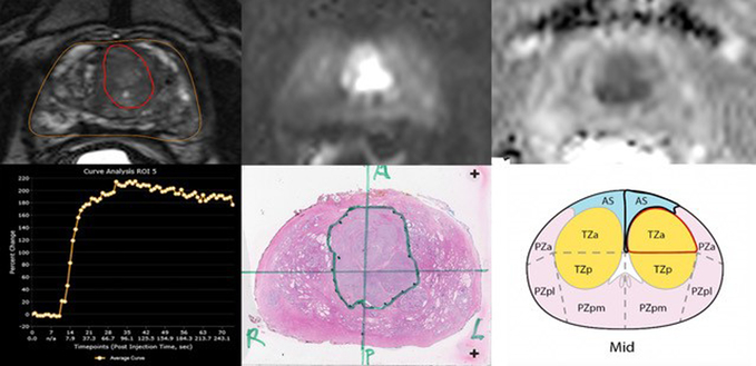
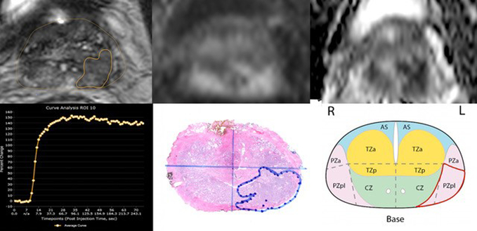
Purpose: To evaluate 3T mpMRI characteristics of transition zone and peripheral zone index prostate cancer lesions stratified by Gleason Score and PI-RADSv2 with whole mount histopathology correlation.
Methods: An institution review board-approved, HIPAA-compliant single-arm observational study of 425 consecutive men with 3T mpMRI prior to radical prostatectomy from December 2009 to October 2016 was performed. A genitourinary radiologist and a genitourinary pathologist matched all lesions detected on whole mount histopathology with lesions concordant for size and location on 3T mpMRI. Differences in clinical, MRI parameters, and histopathology between transition zone and peripheral zone were determined and analyzed with χ2 and Mann-Whitney U test. AUC was measured.
Results: 3T mpMRI detected 248/323 (76.7%) index lesions in peripheral zone and 75/323 (23.2%) in transition zone. Transition zone prostate cancer had higher median prostate-specific antigen (p = 0.001), larger tumor on 3T mpMRI (p = 0.001), lower proportions of PI-RADSv2 category 4 and 5 (p < 0.001), and lower pathological stage (p = 0.055) compared to peripheral zone prostate cancer. No significant differences were detected in prostate-specific antigen density, preoperative biopsy, and pathology Gleason Scores. After adjusting for significant variables from univariate analysis including prostate volume, tumor volume, prostate-specific antigen, PI-RADSv2 category, AUC for predicting clinically significant tumor in transition zone and peripheral zone were 0.80 and 0.72, respectively (p = 0.36).
Conclusions: The diagnostic performance of PI-RADSv2 for clinically significant transition and peripheral zone prostate cancer was similar. However, there was a lower portion of PI-RADSv2 4 and 5 lesions in transition zone compared to peripheral zone.
Keywords: Gleason score; Multiparametric magnetic resonance imaging; PI-RADSv2; Prostate cancer.
Conflict of interest statement
Figures


References
-
- Key statistics for prostate cancer. (2015). http://www.cancer.org/cancer/prostatecancer/detailedguide/prostate-cance.... Accessed 4/29/2016 2016
-
- Early Detection of Prostate Cancer: AUA Guidelines. https://www.auanet.org/education/guidelines/prostate-cancer-detection.cfm. Accessed 4/28/2016 2016
-
- Engelbrecht MR, Huisman HJ, Laheij RJ, Jager GJ, van Leenders GJ, Hulsbergen-Van De Kaa CA, de la Rosette JJ, Blickman JG, Barentsz JO (2003) Discrimination of prostate cancer from normal peripheral zone and central gland tissue by using dynamic contrast-enhanced MR imaging. Radiology 229 (1):248–254. doi:10.1148/radiol.2291020200 - DOI - PubMed
-
- Le JD, Tan N, Shkolyar E, Lu DY, Kwan L, Marks LS, Huang J, Margolis DJ, Raman SS, Reiter RE (2015) Multifocality and prostate cancer detection by multiparametric magnetic resonance imaging: correlation with whole-mount histopathology. Eur Urol 67 (3):569–576. doi:10.1016/j.eururo.2014.08.079 - DOI - PubMed
Publication types
MeSH terms
Substances
Grants and funding
LinkOut - more resources
Full Text Sources
Other Literature Sources
Medical
Research Materials
Miscellaneous
