

Extracellular vesicles generated by placental tissues ex vivo: A transport system for immune mediators and growth factors
- PMID: 29726582
- PMCID: PMC6021205
- DOI: 10.1111/aji.12860
Extracellular vesicles generated by placental tissues ex vivo: A transport system for immune mediators and growth factors
Abstract
Problem: To study the mechanisms of placenta function and the role of extracellular vesicles (EVs) in pregnancy, it is necessary to develop an ex vivo system that retains placental cytoarchitecture and the primary metabolic aspects, in particular the release of EVs and soluble factors. Here, we developed such a system and investigated the pattern of secretion of cytokines, growth factors, and extracellular vesicles by placental villous and amnion tissues ex vivo.
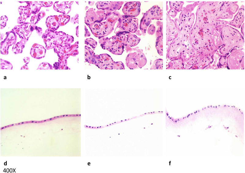
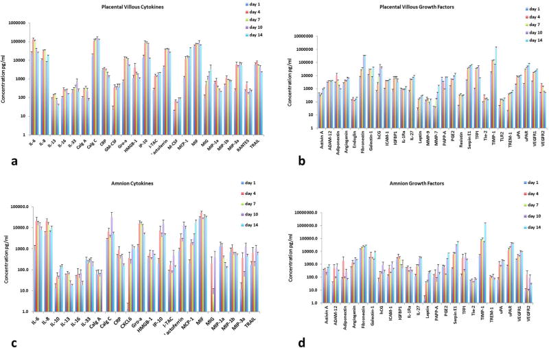
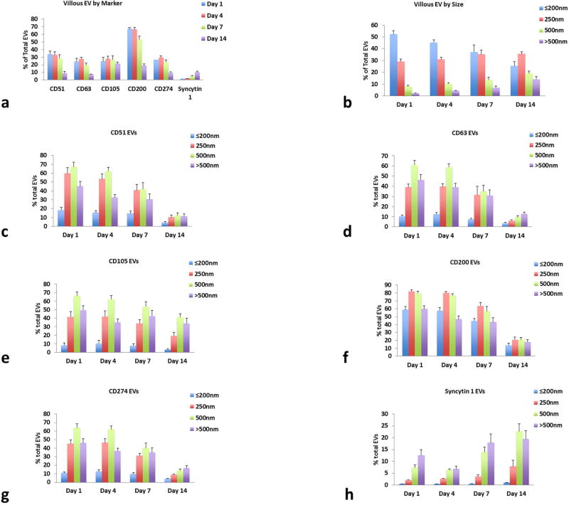
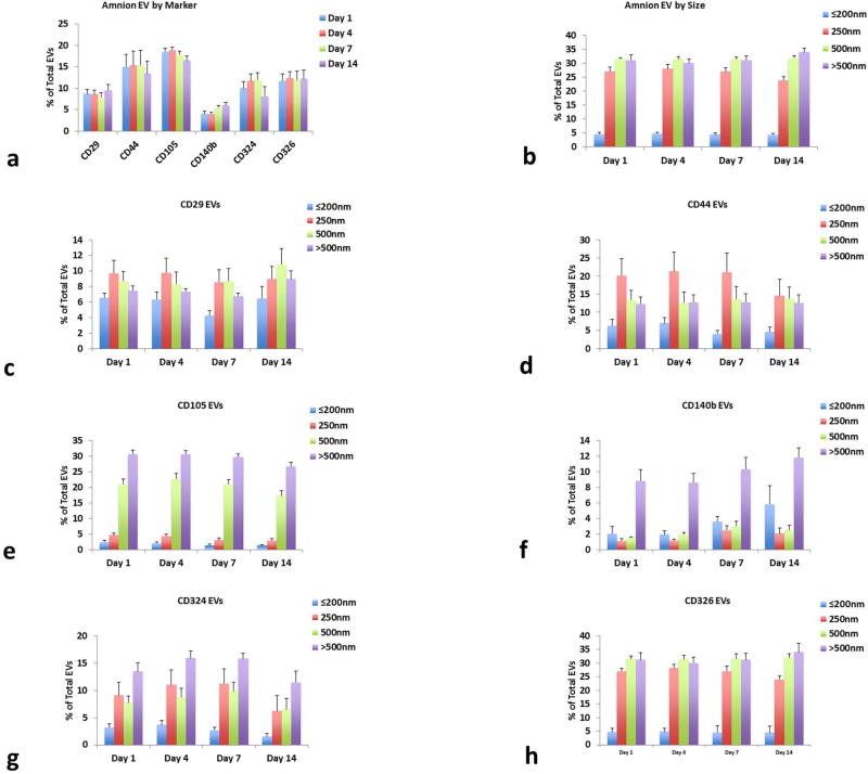
Methods of study: Placental villous and amnion explants were cultured for 2 weeks at the air/liquid interface and their morphology and the released cytokines and EVs were analyzed. Cytokines were analyzed with multiplexed bead assays, and individual EVs were analyzed with recently developed techniques that involved EV capture with magnetic nanoparticles coupled to anti-EV antibodies and flow cytometry.
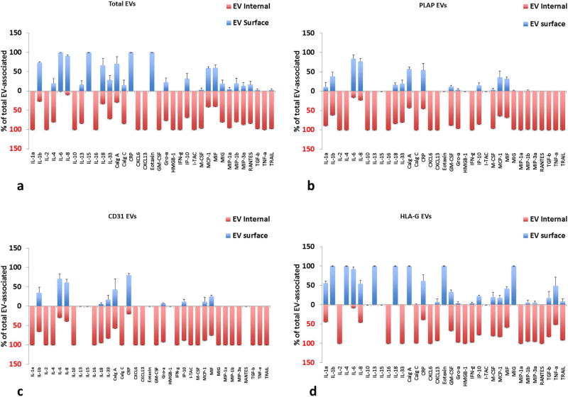
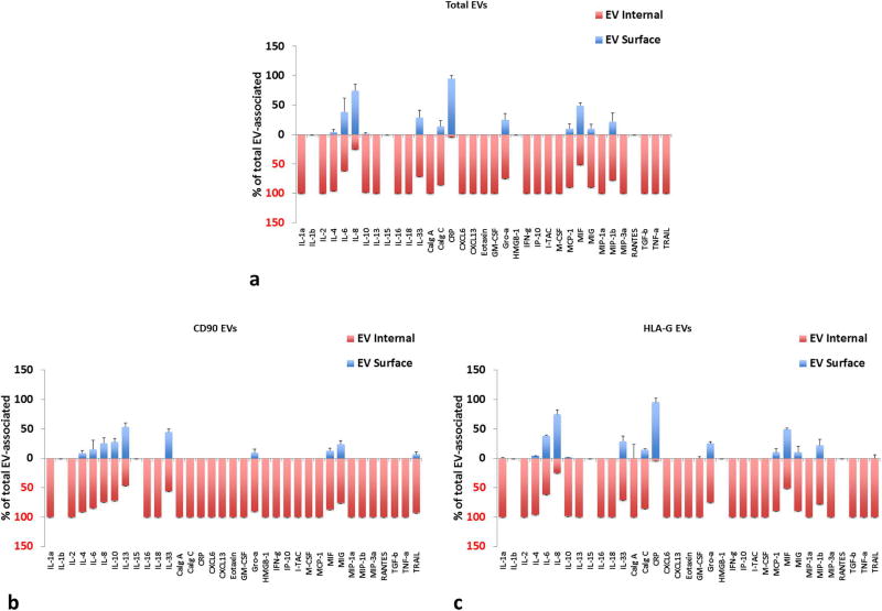
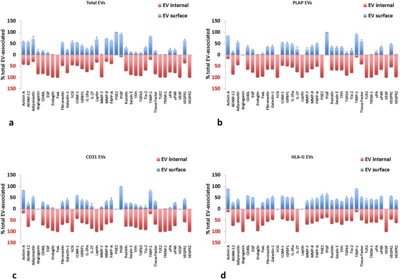
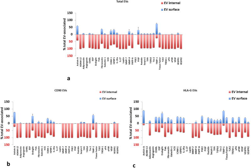
Results: Ex vivo tissues (i) remained viable and preserved their cytoarchitecture; (ii) maintained secretion of cytokines and growth factors; (iii) released EVs of syncytiotrophoblast and amnion epithelial cell origins that contain cytokines and growth factors.
Conclusion: A system of ex vivo placental villous and amnion tissues can be used as an adequate model to study placenta metabolic activity in normal and complicated pregnancies, in particular to characterize EVs by their surface markers and by encapsulated proteins. Establishment and benchmarking the placenta ex vivo system may provide new insight in the functional status of this organ in various placental disorders, particularly regarding the release of EVs and cytokines. Such EVs may have a prognostic value for pregnancy complications.
Keywords: 3D cultures; amnion; cytokine; growth factors; pregnancy; syncytiotrophoblast.
Published 2018. This article is a U.S. Government work and is in the public domain in the USA.
Conflict of interest statement
The authors declare no conflict of interests.
Figures
Similar articles
-
Neuropilin-1 is uniquely expressed on small syncytiotrophoblast extracellular vesicles but not on medium/large vesicles from preeclampsia and normal placentae.Biochem Biophys Res Commun. 2022 Sep 3;619:151-158. doi: 10.1016/j.bbrc.2022.06.041. Epub 2022 Jun 21. Biochem Biophys Res Commun. 2022. PMID: 35760012
-
Reduced placental protein 13 (PP13) in placental derived syncytiotrophoblast extracellular vesicles in preeclampsia - A novel tool to study the impaired cargo transmission of the placenta to the maternal organs.Placenta. 2018 Jun;66:17-25. doi: 10.1016/j.placenta.2018.04.013. Epub 2018 Apr 25. Placenta. 2018. PMID: 29884298
-
Placental Syncytiotrophoblast-Derived Extracellular Vesicles Carry Active NEP (Neprilysin) and Are Increased in Preeclampsia.Hypertension. 2019 May;73(5):1112-1119. doi: 10.1161/HYPERTENSIONAHA.119.12707. Hypertension. 2019. PMID: 30929513
-
Placental small extracellular vesicles: Current questions and investigative opportunities.Placenta. 2020 Dec;102:34-38. doi: 10.1016/j.placenta.2020.03.002. Epub 2020 Mar 10. Placenta. 2020. PMID: 33218576 Free PMC article. Review.
-
Preeclampsia and syncytiotrophoblast membrane extracellular vesicles (STB-EVs).Clin Sci (Lond). 2022 Dec 22;136(24):1793-1807. doi: 10.1042/CS20220149. Clin Sci (Lond). 2022. PMID: 36511102 Free PMC article. Review.
Cited by
-
Unfolding the role of placental-derived Extracellular Vesicles in Pregnancy: From homeostasis to pathophysiology.Front Cell Dev Biol. 2022 Nov 21;10:1060850. doi: 10.3389/fcell.2022.1060850. eCollection 2022. Front Cell Dev Biol. 2022. PMID: 36478738 Free PMC article. Review.
-
Downregulation of Ribosomal Contents and Kinase Activities Is Associated with the Inhibitive Effect on the Growth of Group B Streptococcus Induced by Placental Extracellular Vesicles.Biology (Basel). 2021 Jul 14;10(7):664. doi: 10.3390/biology10070664. Biology (Basel). 2021. PMID: 34356519 Free PMC article.
-
Being pregnant in the COVID-19 pandemic: Effects on the placenta in all aspects.J Med Virol. 2021 May;93(5):2769-2773. doi: 10.1002/jmv.26857. Epub 2021 Feb 15. J Med Virol. 2021. PMID: 33559937 Free PMC article. Review.
-
Antiviral properties of placental growth factors: A novel therapeutic approach for COVID-19 treatment.Placenta. 2020 Sep 15;99:117-130. doi: 10.1016/j.placenta.2020.07.033. Epub 2020 Aug 6. Placenta. 2020. PMID: 32798764 Free PMC article. Review.
-
Are there foetal extracellular vesicles in maternal blood? Prospects for diagnostic biomarker discovery.J Mol Med (Berl). 2023 Feb;101(1-2):65-81. doi: 10.1007/s00109-022-02278-0. Epub 2022 Dec 20. J Mol Med (Berl). 2023. PMID: 36538060 Free PMC article. Review.
References
-
- Desoye G, Shafrir E. Placental metabolism and its regulation in health and diabetes. Mol Aspects Med. 1994;15:505–682. - PubMed
-
- Freemark M. Placental Hormones and the Control of Fetal Growth. The Journal of Clinical Endocrinology & Metabolism. 2010;95:2054–2057. - PubMed
-
- Newbern D, Freemark M. Placental hormones and the control of maternal metabolism and fetal growth. Curr Opin Endocrinol Diabetes Obes. 2011;18:409–416. - PubMed
Publication types
MeSH terms
Substances
Grants and funding
LinkOut - more resources
Full Text Sources
Other Literature Sources
Research Materials
