Randomized Controlled Trial
. 2018 Aug;46(8):e779-e787.
doi: 10.1097/CCM.0000000000003190.

Blue Light Enhances Bacterial Clearance and Reduces Organ Injury During Sepsis

Affiliations
Randomized Controlled Trial

Blue Light Enhances Bacterial Clearance and Reduces Organ Injury During Sepsis

Anthony J Lewis et al. Crit Care Med. 2018 Aug.

Abstract

Objectives: The physiology of nearly all mammalian organisms are entrained by light and exhibit circadian rhythm. The data derived from animal studies show that light influences immunity, and these neurophysiologic pathways are maximally entrained by the blue spectrum. Here, we hypothesize that bright blue light reduces acute kidney injury by comparison with either bright red or standard, white fluorescent light in mice subjected to sepsis. To further translational relevance, we performed a pilot clinical trial of blue light therapy in human subjects with appendicitis.

Design: Laboratory animal research, pilot human feasibility trial.

Setting: University basic science laboratory and tertiary care hospital.

Subjects: Male C57BL/6J mice, adult (> 17 yr) patients with acute appendicitis.

Interventions: Mice underwent cecal ligation and puncture and were randomly assigned to a 24-hour photoperiod of bright blue, bright red, or ambient white fluorescent light. Subjects with appendicitis were randomized to receive postoperatively standard care or standard care plus high-illuminance blue light.

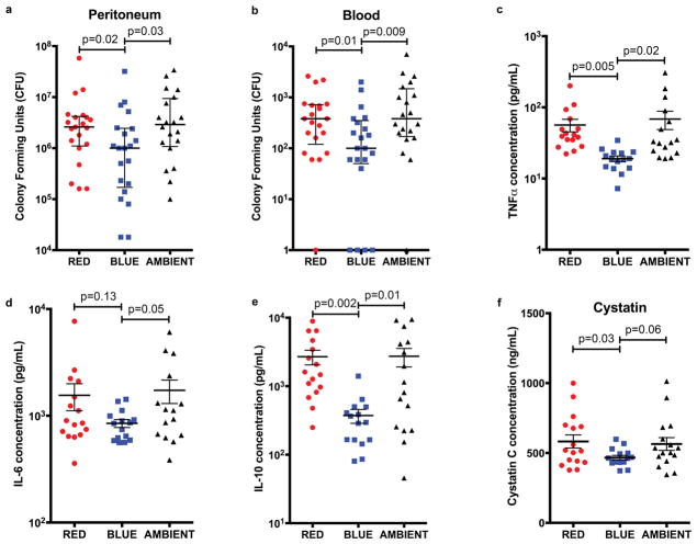
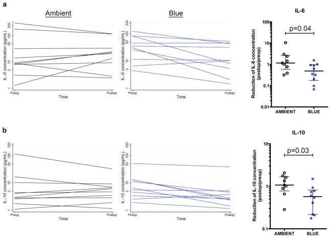
Measurements and main results: Exposure to bright blue light enhanced bacterial clearance from the peritoneum, reduced bacteremia and systemic inflammation, and attenuated the degree of acute kidney injury. The mechanism involved an elevation in cholinergic tone that augmented tissue expression of the nuclear orphan receptor REV-ERBα and occurred independent of alterations in melatonin or corticosterone concentrations. Clinically, exposure to blue light after appendectomy was feasible and reduced serum interleukin-6 and interleukin-10 concentrations.

Conclusions: Modifying the spectrum of light may offer therapeutic utility in sepsis.


Figures

Figure 1
Figure 1. Blue light enhances control of the infection and reduces systemic inflammation and organ injury during CLP sepsis
Mice underwent CLP and then were exposed to red, blue, or ambient light for 24 hours. a and b, Peritoneal lavage fluid and whole blood were cultured for bacteria (18 total mice per group for all 5 experiments combined). bar = median c–f, Serum was analyzed for c, Cystatin C d, TNFα, e, IL-6, and f, IL-10 concentration (16 total mice per group for all 4 experiments combined) mean±sem.
Figure 2
Figure 2. Blue light functions through a cholinergic pathway to attenuate inflammation and organ injury during CLP sepsis
a–c, Mice underwent laparotomy and implantation of a DSI HD-X11 telemetry device. After 24 hours of recovery, mice underwent CLP and were exposed to a 24 hour photoperiod of high illuminance blue or red spectrum light and monitored for 24 hours. Heart rate (HR) was continuously monitored. LF/HF ratio was calculated as a parameter of sympathetic tone and LF as a parameter of parasympathetic tone (57, 58) Each point is a single measurement of a single mouse at a single timepoint. Lines and colored areas represent a fractional polynomial estimate of the mean ± 95% confidence interval of each parameter for each cohort of red (n=8) and blue (n=8) light exposed mice. d, Mice underwent CLP and were randomized to receive α-bungarotoxin (1μg/kg, i.p.) or equivolume vehicle and then exposure to a 24 hour photoperiod of either high illuminance red or blue spectrum light. Serum was analyzed for TNFα, IL-6, IL-10 and Cystatin C concentrations (10 total mice per group for all 3 experiments combined). mean±sem e, Mice underwent CLP and then received either nicotine (1mg/kg, i.p.) or equivolume control. After 24 hours serum was analyzed for TNFα, IL-6, IL-10 and Cystatin C concentrations (9 total mice per group for all 3 experiments combined). mean±sem
Figure 3
Figure 3. Blue light induces the nuclear receptor REV-ERBα in reducing the inflammation and organ injury of sepsis
a, Mice underwent CLP and were exposed to red, blue, or ambient light for 8 hours, at which time point organs were harvested. Tissue expression of REV-ERBα and tubulin was analyzed by immunoblot. (4 mice total in each group). Corresponding densitometry blots compare splenic REV-ERBα expression from septic mice exposed to red, blue, or ambient light. b, Mice underwent CLP and after 4 hours were administered SR9009 (5mg/kg, i.p.). After a total of 24 hours, serum was harvested and assayed for TNFα, IL-6, IL-10 and Cystatin C concentrations. mean±sem (n=8 mice per group) c, Wild type and heterozygous REV-ERBα+/ − mice underwent CLP and were exposed to either red or blue light for 24 hours, at which point serum was harvested and assayed for TNFα, IL-6, IL-10 and Cystatin C concentrations. mean±sem (n=7 mice per group). d, Mice were randomized to receive nicotine (0.4 or 2.0 mg/kg, i.p.) or equivolume normal (0.9%) saline. After 8 hours the organs were harvested, and the tissue expression of REV-ERBα and tubulin was analyzed by immunoblot. (4 mice total in each group). Corresponding densitometry blots compare splenic REV-ERBα expression from septic mice administered normal (0.9%) saline, nicotine (0.4 mg/kg) and nicotine (2.0 mg/kg).
Figure 4
Figure 4. Blue light reduces systemic serum cytokine concentrations in humans with appendicitis
Human subjects with appendicitis were exposed postoperatively to either blue light or ambient light for 18–24 hours. Serum was harvested preoperatively and 24 hours postoperatively and analyzed for a, IL-6 concentration and b, IL-10 concentration. Panel graphs depict serum IL-6 and IL-10 concentrations for each subject at time of admission preoperatively and at approximately 18 hours postoperatively. Corresponding box plots depict median and interquartile range of fold reduction in IL-6 and IL-10, comparing postoperative to preoperative concentrations. (n=10 subjects per group)

Comment in

References

    1. Nelson RJ. Seasonal immune function and sickness responses. Trends in immunology. 2004;25(4):187–192. - PubMed
    1. Prendergast BJ, Kampf-Lassin A, Yee JR, et al. Winter day lengths enhance T lymphocyte phenotypes, inhibit cytokine responses, and attenuate behavioral symptoms of infection in laboratory rats. Brain, behavior, and immunity. 2007;21(8):1096–1108. - PMC - PubMed
    1. Haldar C, Ahmad R. Photoimmunomodulation and melatonin. Journal of photochemistry and photobiology B, Biology. 2010;98(2):107–117. - PubMed
    1. Prendergast BJ. Behavioral tolerance to endotoxin is enhanced by adaptation to winter photoperiods. Psychoneuroendocrinology. 2008;33(4):540–545. - PMC - PubMed
    1. Scheiermann C, Kunisaki Y, Lucas D, et al. Adrenergic nerves govern circadian leukocyte recruitment to tissues. Immunity. 2012;37(2):290–301. - PMC - PubMed

Publication types