. 2018 May;83(5):1027-1031.
doi: 10.1002/ana.25234. Epub 2018 May 4.

Subthalamic nucleus deep brain stimulation evokes resonant neural activity

Affiliations

Subthalamic nucleus deep brain stimulation evokes resonant neural activity

Nicholas C Sinclair et al. Ann Neurol. 2018 May.

Abstract

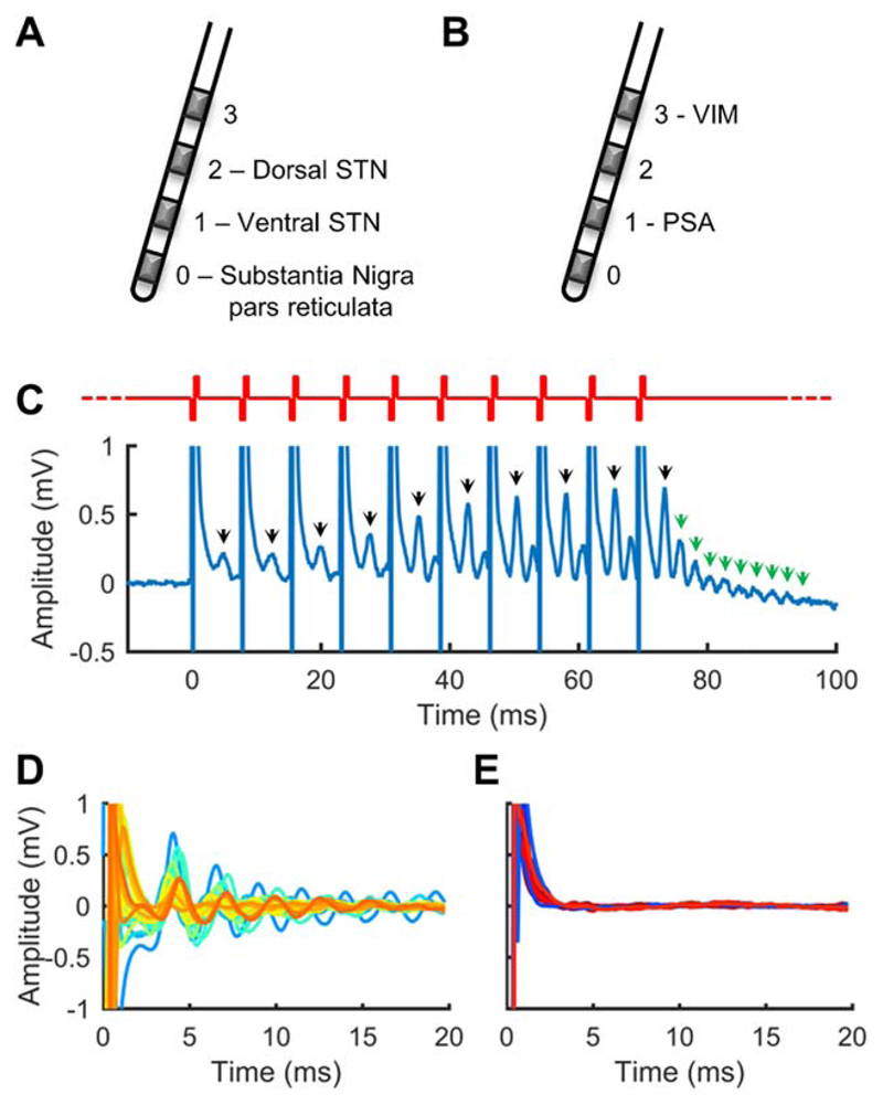
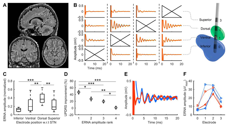
Deep brain stimulation (DBS) is a rapidly expanding treatment for neurological and psychiatric conditions; however, a target-specific biomarker is required to optimize therapy. Here, we show that DBS evokes a large-amplitude resonant neural response focally in the subthalamic nucleus. This response is greatest in the dorsal region (the clinically optimal stimulation target for Parkinson disease), coincides with improved clinical performance, is chronically recordable, and is present under general anesthesia. These features make it a readily utilizable electrophysiological signal that could potentially be used for guiding electrode implantation surgery and tailoring DBS therapy to improve patient outcomes. Ann Neurol 2018;83:1027-1031.


Conflict of interest statement

Potential Conflicts of Interest

Nothing to report.

Figures

Figure 1
Figure 1. Subthalamic nucleus region deep brain stimulation evokes resonant neural activity.
(A) Typical Parkinson disease (PD) subthalamic nucleus (STN) target electrode positions. (B) Typical essential tremor posterior subthalamic area (PSA)/ventral intermediate nucleus (VIM) target electrode positions. (C) Resonant neural activity evoked by a burst of stimulation applied to an electrode in the STN of a PD patient. Bursts comprised 10 pulses delivered at 130Hz (red waveform). Black arrows indicate a peak observable between pulses. Green arrows indicate resonant peaks observable at the end of the burst. Recording electrode: E1; stimulated electrode: E2. (D) Evoked responses from 27 STNs (colors represent different nuclei). Recording electrode: E1; stimulated electrode: E2. (E) Evoked responses from 10 PSAs (blue traces; recording electrode: E0; stimulated electrode: E1) and 8 VIMs (red traces; recording electrode: E2; stimulated electrode: E3). Y-axis is as per D.
Figure 2
Figure 2. Evoked resonant neural activity (ERNA) positional variation, clinical performance and presence under anesthesia.
(A) Example sagittal, coronal, and axial merged magnetic resonance imaging and computed tomography scans used to classify electrode positions. The central hyperintense voxels correspond to the implanted electrodes. (B) End-of-burst ERNA resulting from each electrode being stimulated in the right subthalamic nucleus (STN) of 1 subject. A 3-dimensional reconstruction for the same subject (green: STN; blue: substantia nigra) illustrates the electrode positions. Crossed axes indicate the stimulated electrode, with dashed lines separating each stimulation condition. (C) Normalized ERNA amplitude variation with electrode position across Parkinson disease patients in whom all electrodes were stimulated (20 hemispheres; box: 25th–75th percentiles; line: median; whiskers: range; w.r.t: with respect to). (D) Mean Unified Parkinson’s Disease Rating Scale (UPDRS) improvement from stimulation after ranking electrodes within each hemisphere according to the amplitude of ERNA measured (rank 1: largest ERNA; bars: standard error). Results from 10 PD patients tested post-surgery (20 hemispheres). (E) ERNA recorded in PD09 at electrode implantation (blue) and under general anesthesia 560 days postoperatively (red). (F) ERNA variation across the electrode array at implantation (blue) and 560 days postoperatively (red; solid: left STN; dotted: right STN). *p< 0.05, **p< 0.01, ***p ≤ 0.001.

References

    1. Montgomery EB., Jr . Deep brain stimulation programming: mechanisms, principles, and practice. Oxford, UK: University Press; 2016.
    1. Denys D, Feenstra M, Schuurman R. Deep brain stimulation: a new frontier in psychiatry. Berlin/Heidelberg, Germany: Springer Science + Business Media; 2012.
    1. McDermott H. Neurobionics: the biomedical engineering of neural prostheses. Hoboken, NJ: John Wiley & Sons; 2016. Neurobionics: treatments for disorders of the central nervous system; pp. 213–230.
    1. Pereira EA, Green AL, Nandi D, Aziz TZ. Deep brain stimulation: indications and evidence. Expert Rev Med Devices. 2007;4:591–603. - PubMed
    1. Diamond A, Jankovic J. The effect of deep brain stimulation on quality of life in movement disorders. J Neurol Neurosurg Psychiatry. 2005;76:1188–1193. - PMC - PubMed

Publication types