. 2018 May 3;22(5):769-778.e4.
doi: 10.1016/j.stem.2018.04.001.

Fasting Activates Fatty Acid Oxidation to Enhance Intestinal Stem Cell Function during Homeostasis and Aging

Affiliations

Fasting Activates Fatty Acid Oxidation to Enhance Intestinal Stem Cell Function during Homeostasis and Aging

Maria M Mihaylova et al. Cell Stem Cell. .

Abstract

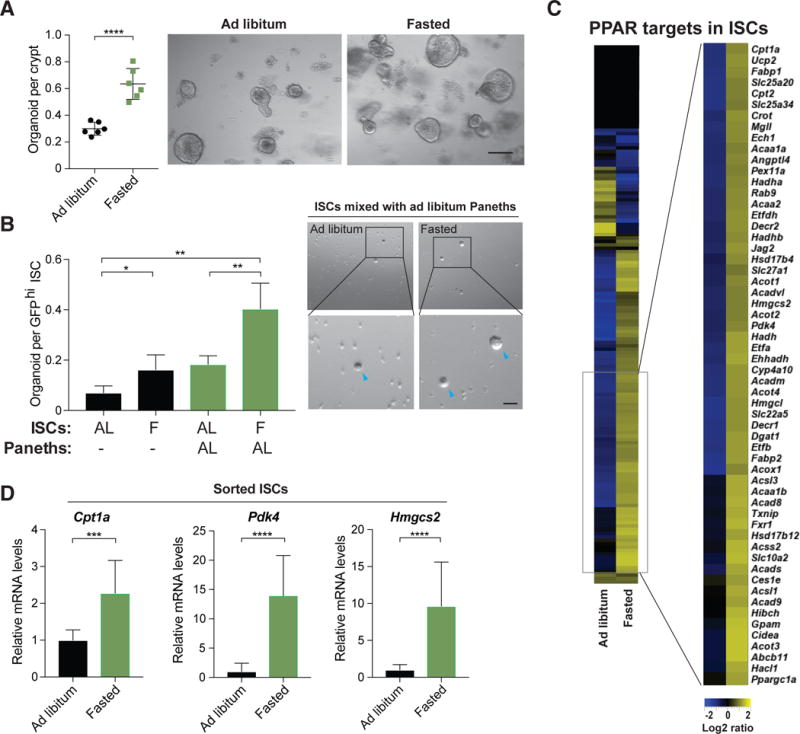
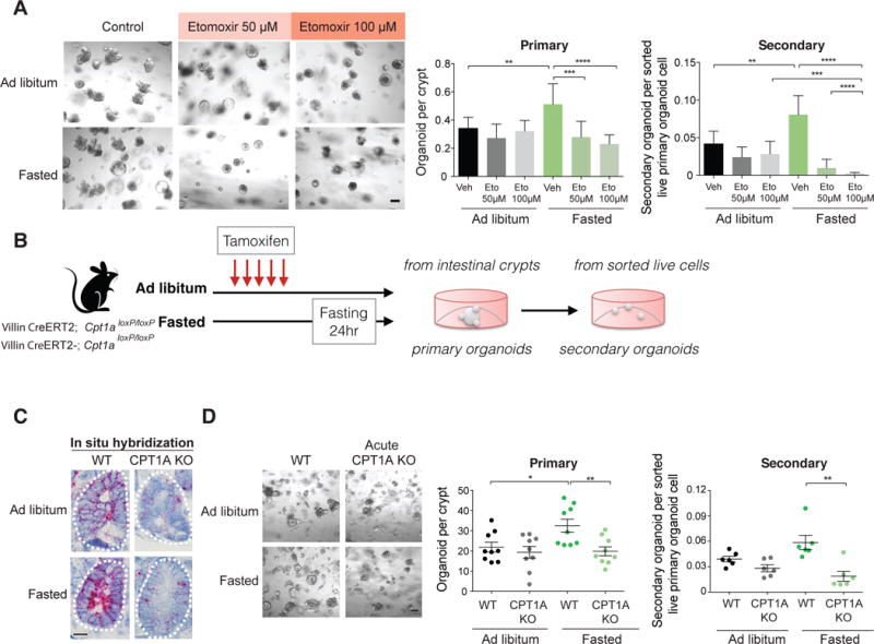
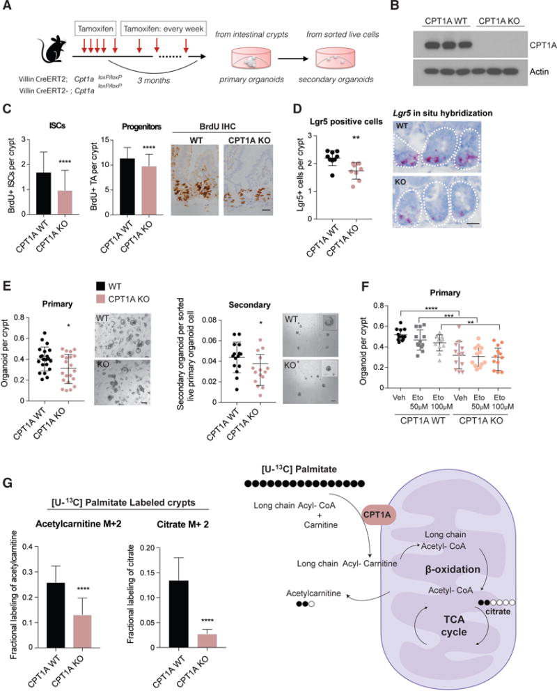
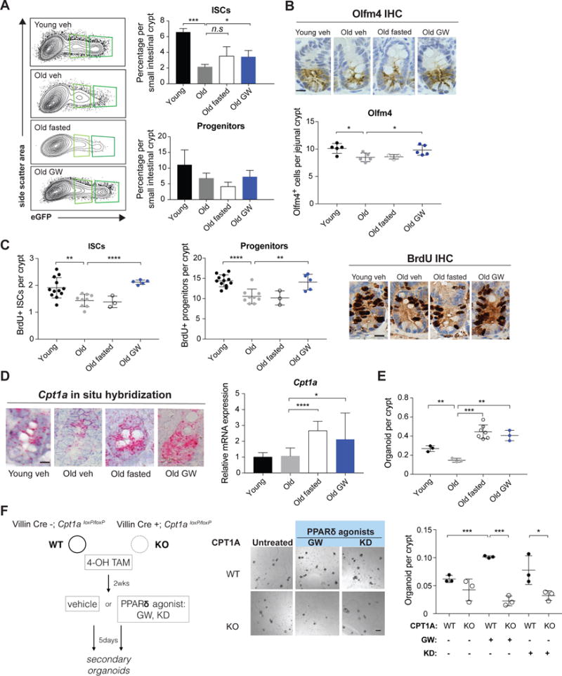
Diet has a profound effect on tissue regeneration in diverse organisms, and low caloric states such as intermittent fasting have beneficial effects on organismal health and age-associated loss of tissue function. The role of adult stem and progenitor cells in responding to short-term fasting and whether such responses improve regeneration are not well studied. Here we show that a 24 hr fast augments intestinal stem cell (ISC) function in young and aged mice by inducing a fatty acid oxidation (FAO) program and that pharmacological activation of this program mimics many effects of fasting. Acute genetic disruption of Cpt1a, the rate-limiting enzyme in FAO, abrogates ISC-enhancing effects of fasting, but long-term Cpt1a deletion decreases ISC numbers and function, implicating a role for FAO in ISC maintenance. These findings highlight a role for FAO in mediating pro-regenerative effects of fasting in intestinal biology, and they may represent a viable strategy for enhancing intestinal regeneration.

Keywords: aging; fasting; fatty acid oxidation; intestinal stem cells; intestine; metabolism; mitochondrial metabolism; stem cells.


Conflict of interest statement

DECLARATION OF INTERESTS

The authors M.M.M., D.M.S. and Ö.H.Y. have filed a patent related to these findings. The authors declare no competing interests.

Figures

Figure 1
Figure 1. Fasting induces fatty acid oxidation and improves intestinal stem cell function
(A) Organoid frequency of crypts from 10-12 week old ad libitum or 24-hour fasted mice. Representative images of crypt culture from each condition. n=6 mice per group. (B) ISCs from 24-hour fasted animals (F) had significantly enhanced organoid-forming capacity when mixed with age matched Paneth cells from ad libitum (AL) mice. Blue arrowheads indicate organoids. n=3 mice per group. (C) Heat map of RNA-Seq analysis showing log2-transformed relative expression of PPAR target genes in ISCs (Lgr5-GFPhi) from AL or 24 hour fasted mice. (D) qRT-PCR analysis confirming PPAR target gene induction in ISCs (Lgr5-GFPhi) following a 24 hour fast. β actin was used as a housekeeping gene. n=7 mice. Data are mean ± s.d. *p < 0.05, **p < 0.01, ***p < 0.001, ****p<0.0005 by Student’s t-test, unpaired. Scale bars, 20 μm (A) and 100 μm (B). See also Figure S1.
Figure 2
Figure 2. Acute ablation of CPT1A abrogates the fasting-induced increase in crypt organoid formation
(A) Administration of etomoxir in culture blunted the effects of 24 hour fasting on organoid formation. Representative images: day 5 primary. n=3 mice. (B) Schematic of acute CPT1A ablation in the intestine. (C) Representative images of in situ hybridization of Cpt1a mRNA levels (red) in crypts of ad libitum or 24 hour fasted wild-type (WT) and CPT1A knockout (CPT1A KO) mice. See also Figures S2E-G. n=3 mice. (D) Deletion of Cpt1a reduced primary and secondary organoid formation of crypts from 24 hour fasted mice. Representative images: day 3 primary organoids. n=3 mice. Data are mean ± s.d. *p < 0.05, **p < 0.01, ***p < 0.001, ****p<0.0005 by Student’s t-test, unpaired. Scale bars, 20 μm (A, C and D). See also Figure S2.
Figure 3
Figure 3. Long-term ablation of CPT1A diminishes ISC numbers and function
(A) Schematic of long-term (3 months) ablation of CPT1A in the intestine. (B) Western blot analysis of CPT1A protein levels in intestinal crypts from WT and long-term deleted CPT1A KO mice. (C) BrdU+ ISCs and progenitor cells in WT and CPT1A KO mice. (D) Long-term deletion of intestinal CPT1A decreased Lgr5+ stem cell numbers. n= 3 mice per group. Representative images of Lgr5+ cells by in situ hybridization (red). (E) Long-term loss of CPT1A compromised organoid-forming capacity in primary and secondary cultures. n= 5 to 7 mice per group, (F) In vitro etomoxir treatment did not further reduce the clonogenic ability of CPT1A KO crypts. n=4 mice. (G) Long-term deletion of CPT1A significantly reduced the contribution of [U-13C] palmitate to acetylcarnitine and citrate. n=6 mice per group. Data are mean ± s.d. *p < 0.05, **p < 0.01, ***p < 0.001, ****p<0.0005. Student’s t-test, unpaired. Scale bars, 50 μm (C and D), 20 μm primary and 100 μm secondary (E). Also see figure S3.
Figure 4
Figure 4. Physiological and pharmacological activation of PPAR delta and subsequent increase of FAO boost intestinal stem cell function in aged animals
(A) FACS analysis of ISC (Lgr5-GFPhi) and progenitor cell (Lgr5-GFPlow) frequency in 4 month or 18-22 month old mice treated with either vehicle, GW501516 (GW, 3-4 weeks) or fasted for 24 hours. n=6 for young, n=7 for old, n=6 for old GW, n= 3 for 24 hour fasted mice. (B) Quantification by IHC of Olfm4+ cells in young vehicle treated, old vehicle treated, old fasted and old GW treated animals. n=5 per group. (C) BrdU incorporation in ISCs and progenitors in young and aged animals following a 4-hour pulse . n=11 for young, n=9 for old, n= 3 for old fasted and n=5 for old GW treated mice. (D) GW treatment and fasting strongly induce Cpt1a expression in aged intestinal stem and progenitor cells. Representative images of Cpt1a in situ hybridization (left) and qRT-PCR analysis of Cpt1a expression in sorted ISCs from corresponding treatments (right). n=12 for young, n=12 for old, n= 11 and n=8 for old GW treated mice. (E) Intestinal crypts from aged animals have diminished capacity for organoid formation compared to young controls. GW treatment (3-4 weeks) and 24-hour fasting of old animals significantly boost organoid-forming capacity of crypts relative to controls. n= 3 independent experiments. (F) Deletion of CPT1A prevented the organoid-enhancing effects of GW treatment. Cpt1aloxP/loxP (WT) and Villin-CreERT2; Cpt1aloxP/loxP (KO) organoids were treated with 4-OH tamoxifen in culture to delete CPT1A and subsequently treated with PPARδ agonists GW501516 (GW, 1 M) and KD3010 (KD, 3 M) or vehicle for 5 days. Equal number of live cells from the treated organoids were sorted to ascertain organoid initiation in secondary organoids. Data are mean ± s.d. *p < 0.05, **p < 0.01, ***p < 0.001 by Student’s t-test, unpaired. Scale bars, 20 m (B, C and D) and 100 m (F). Also see figure S4.

Comment in

References

    1. Anders S, Huber W. Differential expression analysis for sequence count data. Genome biology. 2010;11:R106. - PMC - PubMed
    1. Anders S, Pyl PT, Huber W. HTSeq–a Python framework to work with high-throughput sequencing data. Bioinformatics. 2015;31:166–169. - PMC - PubMed
    1. Asfaha S, Hayakawa Y, Muley A, Stokes S, Graham TA, Ericksen RE, Westphalen CB, von Burstin J, Mastracci TL, Worthley DL, et al. Krt19(+)/Lgr5(-) Cells Are Radioresistant Cancer-Initiating Stem Cells in the Colon and Intestine. Cell stem cell. 2015;16:627–638. - PMC - PubMed
    1. Barish GD, Narkar VA, Evans RM. PPAR delta: a dagger in the heart of the metabolic syndrome. The Journal of clinical investigation. 2006;116:590–597. - PMC - PubMed
    1. Barker N, van Es JH, Kuipers J, Kujala P, van den Born M, Cozijnsen M, Haegebarth A, Korving J, Begthel H, Peters PJ, et al. Identification of stem cells in small intestine and colon by marker gene Lgr5. Nature. 2007;449:1003–1007. - PubMed

Publication types