

Stenting the Eustachian tube to treat chronic otitis media - a feasibility study in sheep
- PMID: 29728102
- PMCID: PMC5935938
- DOI: 10.1186/s13005-018-0165-5
Stenting the Eustachian tube to treat chronic otitis media - a feasibility study in sheep
Abstract
Background: Untreated chronic otitis media severely impairs quality of life in affected individuals. Local destruction of the middle ear and subsequent loss of hearing are common sequelae, and currently available treatments provide limited relief. Therefore, the objectives of this study were to evaluate the feasibility of the insertion of a coronary stent from the nasopharynx into the Eustachian tube in-vivo in sheep and to make an initial assessment of its positional stability, tolerance by the animal, and possible tissue reactions.
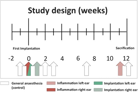
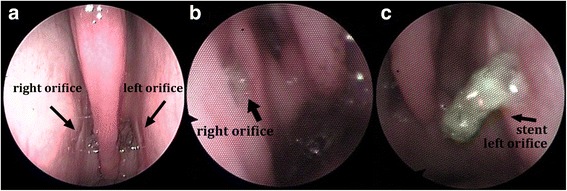
Methods: Bilateral implantation of bare metal cobalt-chrome coronary stents of two sizes was performed endoscopically in three healthy blackface sheep using a nasopharyngeal approach. The postoperative observation period was three months.
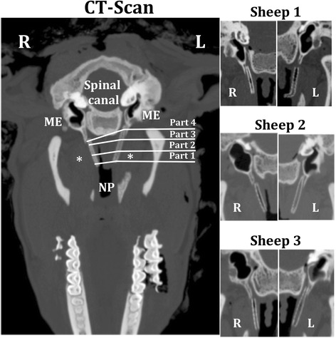
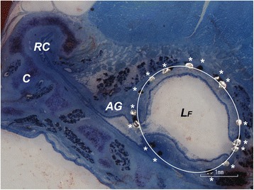
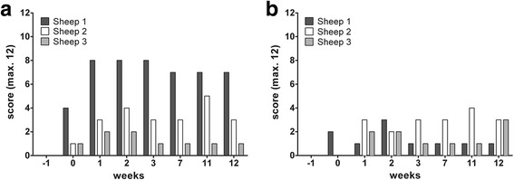
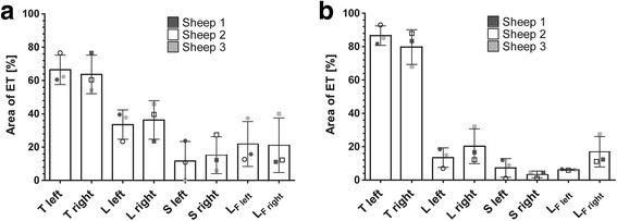
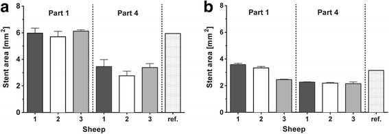
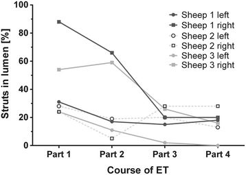
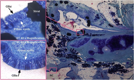
Results: Stent implantation into the Eustachian tube was feasible with no intra- or post-operative complications. Health status of the sheep was unaffected. All stents preserved their cylindrical shape. All shorter stents remained in position and ventilated the middle ear even when partially filled with secretion or tissue. One of the long stents became dislocated toward the nasopharynx. Both of the others remained fixed at the isthmus but appeared to be blocked by tissue or secretion. Tissue overgrowth on top of the struts of all stents resulted in closure of the tissue-lumen interface.
Conclusion: Stenting of the Eustachian tube was successfully transferred from cadaver studies to an in-vivo application without complications. The stent was well tolerated, the middle ears were ventilated, and clearance of the auditory tube appeared possible. For fixation, it seems to be sufficient to place it only in the cartilaginous part of the Eustachian tube.
Keywords: Auditory tube; Middle ear ventilation; Otitis media; Sheep as animal model; Stent; Tissue reaction.
Conflict of interest statement
Ethics approval and consent to participate
The State Office for Consumer Protection and Food Safety, Dept. of Animal Welfare in accordance with the German and European animal welfare legislation approved this study under the number 12/1089.
Competing interests
The authors declare that they have no competing interests.
Publisher’s Note
Springer Nature remains neutral with regard to jurisdictional claims in published maps and institutional affiliations.
Figures
Similar articles
-
Treatment of middle ear ventilation disorders: sheep as animal model for stenting the human Eustachian tube--a cadaver study.PLoS One. 2014 Nov 24;9(11):e113906. doi: 10.1371/journal.pone.0113906. eCollection 2014. PLoS One. 2014. PMID: 25419714 Free PMC article.
-
[Use of NiTi shape memory alloys in the eustachian tube to prevent and treat adhesive middle ear].Zhonghua Er Bi Yan Hou Ke Za Zhi. 2002 Feb;37(1):55-7. Zhonghua Er Bi Yan Hou Ke Za Zhi. 2002. PMID: 12768796 Chinese.
-
Intraluminal three-dimensional optical coherence tomography - a tool for imaging of the Eustachian tube?J Laryngol Otol. 2019 Feb;133(2):87-94. doi: 10.1017/S002221511800230X. Epub 2019 Feb 18. J Laryngol Otol. 2019. PMID: 30773144
-
Update on eustachian tube dysfunction and the patulous eustachian tube.Curr Opin Otolaryngol Head Neck Surg. 2005 Oct;13(5):277-82. doi: 10.1097/01.moo.0000176465.68128.45. Curr Opin Otolaryngol Head Neck Surg. 2005. PMID: 16160520 Review.
-
Anatomy and physiology of eustachian tube and middle ear related to otitis media.J Allergy Clin Immunol. 1988 May;81(5 Pt 2):997-1003. doi: 10.1016/0091-6749(88)90168-6. J Allergy Clin Immunol. 1988. PMID: 3286738 Review.
Cited by
-
Development of an In Vivo Model for Eustachian Tube Dysfunction.Bioengineering (Basel). 2022 Jul 15;9(7):317. doi: 10.3390/bioengineering9070317. Bioengineering (Basel). 2022. PMID: 35877368 Free PMC article.
-
Tapered self-expandable metallic stent optimized for Eustachian tube morphology in a porcine ET model.Sci Rep. 2022 Nov 24;12(1):20290. doi: 10.1038/s41598-022-24615-6. Sci Rep. 2022. PMID: 36434004 Free PMC article.
-
Long-Term Preclinical Evaluation of a Permanent Stent Developed for the Human Eustachian Tube.Bioengineering (Basel). 2024 Jul 25;11(8):755. doi: 10.3390/bioengineering11080755. Bioengineering (Basel). 2024. PMID: 39199713 Free PMC article.
-
Comparative study of optical coherence tomograph and histological images of eustachian tube nasopharyngeal region and adjacent structures in vivo and ex-vivo miniature pigs.Biomed Eng Online. 2023 May 13;22(1):46. doi: 10.1186/s12938-023-01104-z. Biomed Eng Online. 2023. PMID: 37179353 Free PMC article.
-
Investigation of Stent Prototypes for the Eustachian Tube in Human Donor Bodies.Bioengineering (Basel). 2023 Jun 20;10(6):743. doi: 10.3390/bioengineering10060743. Bioengineering (Basel). 2023. PMID: 37370674 Free PMC article.
References
MeSH terms
LinkOut - more resources
Full Text Sources
Other Literature Sources
Medical
