. 2018 Jul 20;123(3):335-341.
doi: 10.1161/CIRCRESAHA.118.313225. Epub 2018 May 4.

CRISPR-Mediated Gene Editing to Assess the Roles of Tet2 and Dnmt3a in Clonal Hematopoiesis and Cardiovascular Disease

Affiliations

CRISPR-Mediated Gene Editing to Assess the Roles of Tet2 and Dnmt3a in Clonal Hematopoiesis and Cardiovascular Disease

Soichi Sano et al. Circ Res. .

Abstract

Rationale: Clonal hematopoiesis has been associated with increased mortality and cardiovascular disease. This condition can arise from somatic mutations in preleukemic driver genes within hematopoietic stem/progenitor cells. Approximately 40 candidate driver genes have been identified, but mutations in only 1 of these genes, TET2 (ten-eleven translocation-2), has been shown to casually contribute to cardiovascular disease in murine models.

Objective: To develop a facile system to evaluate the disease characteristics of different clonal hematopoiesis driver genes using lentivirus vector and CRISPR/Cas9 (clustered regularly interspaced short palindromic repeats/clustered regularly interspaced short palindromic repeat-associated 9) methodology. Using this methodology, evaluate whether Dnmt3a (DNA [cytosine-5]-methyltransferase 3a)-a commonly occurring clonal hematopoiesis driver gene-causally contributes to cardiovascular disease.

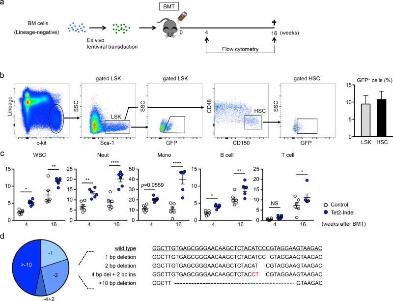
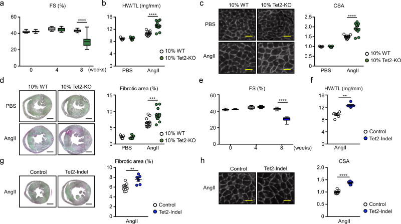
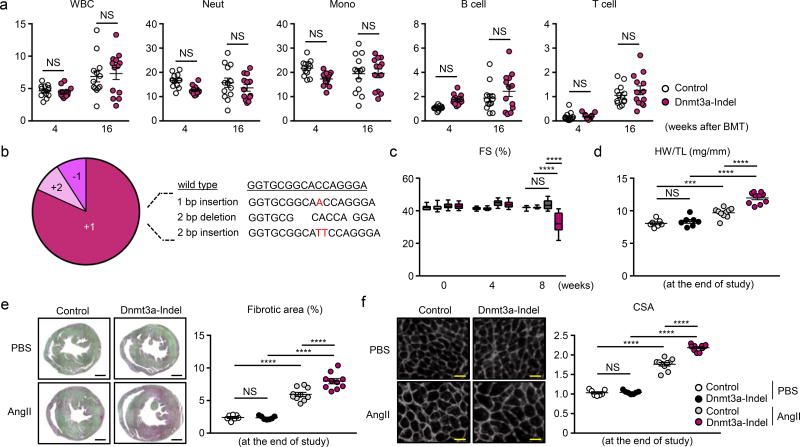
Methods and results: Lentivirus vectors were used to deliver Cas9 and guide RNA to introduce inactivating mutations in Tet2 and Dnmt3a in lineage-negative bone marrow cells. After implantation into lethally irradiated mice, these cells were engrafted and gave rise to labeled blood cell progeny. When challenged with an infusion of Ang II (angiotensin II), mice with inactivating mutations in Tet2 or Dnmt3a displayed greater cardiac hypertrophy, diminished cardiac function, and greater cardiac and renal fibrosis. In comparison with Tet2, inactivation of Dnmt3a did not lead to detectable expansion of the mutant hematopoietic cells during the time course of these experiments. Tet2 inactivation promoted the expression of IL (interleukin) 1β, IL-6, and Ccl5, whereas Dnmt3a inactivation promoted the expression of Cxcl1 (CXC chemokine ligand), Cxcl2, IL-6, and Ccl5 in a lipopolysaccharide-stimulated macrophage cell line.

Conclusions: Experiments using lentivirus vector/CRISPR methodology provided evidence suggesting that inactivating DNMT3A mutations in hematopoietic cells contributes to cardiovascular disease. Comparative analyses showed that inactivation of Tet2 and Dnmt3 was similar in their ability to promote Ang II-induced cardiac dysfunction and renal fibrosis in mice. However, gene-specific actions were indicated by differences in kinetics of hematopoietic stem/progenitor cell expansion and different patterns of inflammatory gene expression.

Keywords: animals; clustered regularly interspaced short palindromic repeats; genetics; heart failure; stem cells.


Figures

Figure 1
Figure 1. CRISPR/Cas9-mediated Tet2 gene disruption confers a competitive advantage to the hematopoietic stem/progenitor cells
a. Bone marrow lineage-negative cells from wild type mice were transduced with lentivirus particles expressing Cas9/eGFP and delivered to lethally irradiated wild type mice. b. Flow cytometry analysis of HSPC transduction by lentivirus. Cells are defined as LSK cells (lineage, c-kit+, Sca-1+) and HSC (CD48, CD150+ in LSK cells). Transduced cells are GFP positive (n=4). c. Flow cytometry analysis of the peripheral blood at 4 and 16 weeks after reconstitution with bone marrow transduced with Tet2-targeted and control (no Tet2 guide RNA) lentivirus vectors. The percentage of GFP+ cells in both experimental groups are shown (n=6 in both Tet2-indel mice and control mice). Statistical analysis was evaluated by two-way repeated measure ANOVA with Sidak’s multiple comparison tests. d. Results of the TA cloning procedure showing that GFP+ peripheral white blood cells harbor edited Tet2 genes. The wild-type Tet2 sequence is shown for reference. *p<0.05, **p<0.01, ****p<0.0001.
Figure 2
Figure 2. Phenotype comparison of Angiotensin-II infusion-induced cardiac dysfunction between conventional competitive BMT model and lenti-CRISPR/Cas9 model
a–d. Data from conventional competitive BMT model. e–h. Data from lenti-CRISPR/Cas9 model. a. Echocardiographic analysis at the indicated time points after AngII infusion (12 mice per group). Statistical analysis was evaluated by two-way repeated measure ANOVA with Sidak’s multiple comparison tests. b. HW adjusted by TL at the end of the study (8 weeks). Statistical analysis was evaluated by two-way ANOVA with Sidak’s multiple comparison tests. c. Representative images and analysis of WGA staining of the heart sections from hearts of 10% KO-BMT mice and 10% WT-BMT mice at the end of the study. Statistical analysis was evaluated by two-way ANOVA with Sidak’s multiple comparison tests. Scale bar: 25µm. d. Representative images and analysis of picrosirius red staining of the heart sections from hearts of 10% KO-BMT mice and 10% WT-BMT mice at the end of the study. Statistical analysis was evaluated by two-way ANOVA with Sidak’s multiple comparison tests. Scale bar: 1mm. In b–d, n=12 for AngII groups and n= 5 for PBS groups were analyzed. e. Echocardiographic analysis at indicated time points after AngII infusion (6 mice per group). Statistical analysis was evaluated by two-way repeated measure ANOVA with Sidak’s multiple comparison tests. f. HW adjusted by TL at the end of the study (8 weeks). Statistical analysis was evaluated by Mann-Whitney U test. g. Representative images and analysis of WGA staining of the heart sections from hearts of Tet2-indel mice and control mice at the end of the study. Scale bar: 1mm. Statistical analysis was evaluated by two-tailed unpaired Student’s t test. h. Representative images and analysis of Picro sirius staining of the heart sections from hearts of Tet2-indel mice and control mice at the end of the study. Scale bar: 25µm. Statistical analysis was evaluated by two-tailed unpaired Student’s t test. In e–h, n=6 per group were analyzed. **p<0.01, ***p<0.001, ****p<0.0001.
Figure 3
Figure 3. CRISPR/Cas9-mediated hematopoietic Dnmt3a gene disruption promotes cardiac dysfunction after AngII infusion
a. Flow cytometry analysis of the peripheral blood at 4 and 16 weeks after bone marrow reconstitution. The percentages of GFP+ cells in both experimental groups are shown (n=13 in both Dnmt3a-indel mice and control mice). Statistical analysis was evaluated by two-way repeated measure ANOVA with Sidak’s multiple comparison tests. b. The result of the TA cloning procedure showing that GFP+ peripheral white blood cells harbors edited Dnmt3a genes. The wild-type Dnmt3a sequence is shown for reference. c. Echocardiographic analysis at indicated time points after AngII infusion (10 mice per group). Statistical analysis was evaluated by two-way repeated measure ANOVA with Tukey’s multiple comparison tests. d. HW adjusted by TL at the end of the study (2 months). Statistical analysis was evaluated by two-way ANOVA with Tukey’s multiple comparison tests. e. Representative images and analysis of picrosirius staining of the heart sections from hearts of Dnmt3a-indel mice and control mice at the end of the study. Scale bar: 1mm. Statistical analysis was evaluated by two-way ANOVA with Tukey’s multiple comparison tests. f. Representative images and analysis of WGA staining of the heart sections from hearts of Dnmt3a-indel mice and control mice at the end of the study. Scale bar: 25µm. Statistical analysis was evaluated by two-way ANOVA with Tukey’s multiple comparison tests. In d–f, n=10 for AngII groups and n=7 for PBS groups were analyzed. ***p<0.001, ****p<0.0001, NS: not significant.
Figure 4
Figure 4. Hematopoietic Dnmt3a loss-of-function enhances cardiac inflammation
a. Western blot analysis revealing decreased in Dnmt3a expression in J774.1 cells treated with lentivirus-mediated Dnmt3a knock out. The lentivirus without sgRNA was used as control. b. Gene expression analysis of WT, Tet2-indel, and Dnmt3a-indel J774.1 cells at 6 hours after stimulation with10 ng/ml LPS. Statistical analysis was evaluated by two-way ANOVA with Tukey’s multiple comparison tests. c. Representative images and analysis of Mac2 staining of the sections of hearts from Dnmt3a-indel mice and control mice at 8 weeks after AngII infusion (n=6 per group). Scale bar: 100µm. Statistical analysis was evaluated by Mann-Whitney U test. d. Gene expression analysis of heart from Dnmt3a-indel and Control mice (n=10 per group) 8 weeks after AngII infusion. Statistical analysis was evaluated by two-tailed unpaired Student’s t test or Mann-Whitney U test. *p<0.05, **p<0.01, ***p<0.001, ****p<0.0001.

Comment in

References

    1. Vijg J. Somatic mutations, genome mosaicism, cancer and aging. Curr Opin Genet Dev. 2014;26:141–9. - PMC - PubMed
    1. Fuster JJ, Walsh K. Somatic Mutations and Clonal Hematopoiesis: Unexpected Potential New Drivers of Age-Related Cardiovascular Disease. Circ Res. 2018;122:523–532. - PMC - PubMed
    1. Steensma DP, Bejar R, Jaiswal S, Lindsley RC, Sekeres MA, Hasserjian RP, Ebert BL. Clonal hematopoiesis of indeterminate potential and its distinction from myelodysplastic syndromes. Blood. 2015;126:9–16. - PMC - PubMed
    1. Jaiswal S, Fontanillas P, Flannick J, Manning A, Grauman PV, Mar BG, Lindsley RC, Mermel CH, Burtt N, Chavez A, Higgins JM, Moltchanov V, Kuo FC, Kluk MJ, Henderson B, Kinnunen L, Koistinen HA, Ladenvall C, Getz G, Correa A, Banahan BF, Gabriel S, Kathiresan S, Stringham HM, McCarthy MI, Boehnke M, Tuomilehto J, Haiman C, Groop L, Atzmon G, Wilson JG, Neuberg D, Altshuler D, Ebert BL. Age-related clonal hematopoiesis associated with adverse outcomes. N Engl J Med. 2014;371:2488–98. - PMC - PubMed
    1. Genovese G, Kahler AK, Handsaker RE, Lindberg J, Rose SA, Bakhoum SF, Chambert K, Mick E, Neale BM, Fromer M, Purcell SM, Svantesson O, Landen M, Hoglund M, Lehmann S, Gabriel SB, Moran JL, Lander ES, Sullivan PF, Sklar P, Gronberg H, Hultman CM, McCarroll SA. Clonal hematopoiesis and blood-cancer risk inferred from blood DNA sequence. N Engl J Med. 2014;371:2477–87. - PMC - PubMed

Publication types

MeSH terms