Review
. 2018 Jun;11(2):201-208.
doi: 10.1007/s12178-018-9475-2.

The medial patellofemoral complex

Affiliations
Review

The medial patellofemoral complex

Alexander E Loeb et al. Curr Rev Musculoskelet Med. 2018 Jun.

Abstract

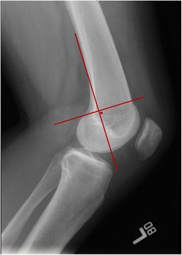
Purpose of review: The purpose of this review is to describe the current understanding of the medial patellofemoral complex, including recent anatomic advances, evaluation of indications for reconstruction with concomitant pathology, and surgical reconstruction techniques.

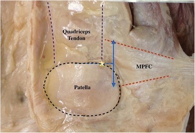
Recent findings: Recent advances in our understanding of MPFC anatomy have found that there are fibers that insert onto the deep quadriceps tendon as well as the patella, thus earning the name "medial patellofemoral complex" to allow for the variability in its anatomy. In MPFC reconstruction, anatomic origin and insertion points and appropriate graft length are critical to prevent overconstraint of the patellofemoral joint. The MPFC is a crucial soft tissue checkrein to lateral patellar translation, and its repair or reconstruction results in good restoration of patellofemoral stability. As our understanding of MPFC anatomy evolves, further studies are needed to apply its relevance in kinematics and surgical applications to its role in maintaining patellar stability.

Keywords: MPFC; MPFL; Medial patellofemoral complex; Medial patellofemoral ligament; Patellar instability.


Conflict of interest statement

Conflict of interest

Both authors declare that they have no conflict of interest.

Human and animal rights and informed consent

All reported studies/experiments with human or animal subjects performed by the authors have been previously published and complied with all applicable ethical standards (including the Helsinki declaration and its amendments, institutional/national research committee standards, and international/national/institutional guidelines).

Figures

Fig. 1
Fig. 1
The anatomy of the MPFC (dashed red line) is shown from the articular surface, with fibers inserting on the patella and the quadriceps tendon (dashed purple line). The midpoint (dashed blue line) is shown at the junction of the medial border of the quadriceps tendon with the patella (yellow star)
Fig. 2.
Fig. 2.
The glide test is used to quantify medial and lateral patellar translation based on patellar quadrants, with two quadrants in each direction typically representing normal motion
Fig. 3
Fig. 3
Axial view of the right knee on T2 fat-suppressed MR imaging demonstrates and effusion and increased lateral translation and tilt after patellar dislocation. A typically bony edema pattern is demonstrated on the medial patella and lateral femoral condyle (dotted yellow arrows). Additionally, injury to the MPFC at the midsubstance is noted (red arrow)
Fig. 4
Fig. 4
Both single-bundle and double-bundled reconstruction of the MPFC have been described. The figure demonstrates an intraoperative example of single-bundle MPFC reconstruction utilizing the docking technique
Fig. 5
Fig. 5
Schottle’s point is demonstrated on a lateral radiograph of a left knee, approximating the femoral origin of the MPFC. This is found anterior to the posterior cortical line, and distal to the posterior medial femoral condyle, and can be utilized intraoperatively to guide femoral tunnel placement during MPFC reconstruction

References

    1. Weber AE, Nathani A, Dines JS, Allen AA, Shubin-Stein BE, Arendt EA, et al. An algorithmic approach to the management of recurrent lateral patellar dislocation. J Bone Jt Surg. 2016;98(5):417–27. 10.2106/JBJS.O.00354. - PubMed
    1. Aframian A, Smith TO, Tennent TD, Cobb JP, Hing CB. Origin and insertion of the medial patellofemoral ligament: a systematic review of anatomy. Knee Surgery, Sport Traumatol Arthrosc. 2016;25:1–18. - PMC - PubMed
    1. Tanaka MJ, Voss A, Fulkerson JP. The anatomic midpoint of the attachment of the medial patellofemoral complex. J Bone Joint Surg Am. 2016;98(14):1199–1205. doi: 10.2106/JBJS.15.01182. - DOI - PubMed
    1. Tanaka MJ. The anatomy of the medial patellofemoral ligament. Sports Med Arthrosc. 2017;25(2):8–11. doi: 10.1097/JSA.0000000000000143. - DOI - PubMed
    1. Tanaka MJ. Variability in the patellar attachment of the medial patellofemoral ligament. Arthrosc - J Arthrosc Relat Surg. 2016;32(8):1667–1670. doi: 10.1016/j.arthro.2016.01.046. - DOI - PubMed

LinkOut - more resources