. 2018 Jun 28:280:11-19.
doi: 10.1016/j.jconrel.2018.04.048. Epub 2018 May 2.

Single Step Double-walled Nanoencapsulation (SSDN)

Affiliations

Single Step Double-walled Nanoencapsulation (SSDN)

Aharon Azagury et al. J Control Release. .

Abstract

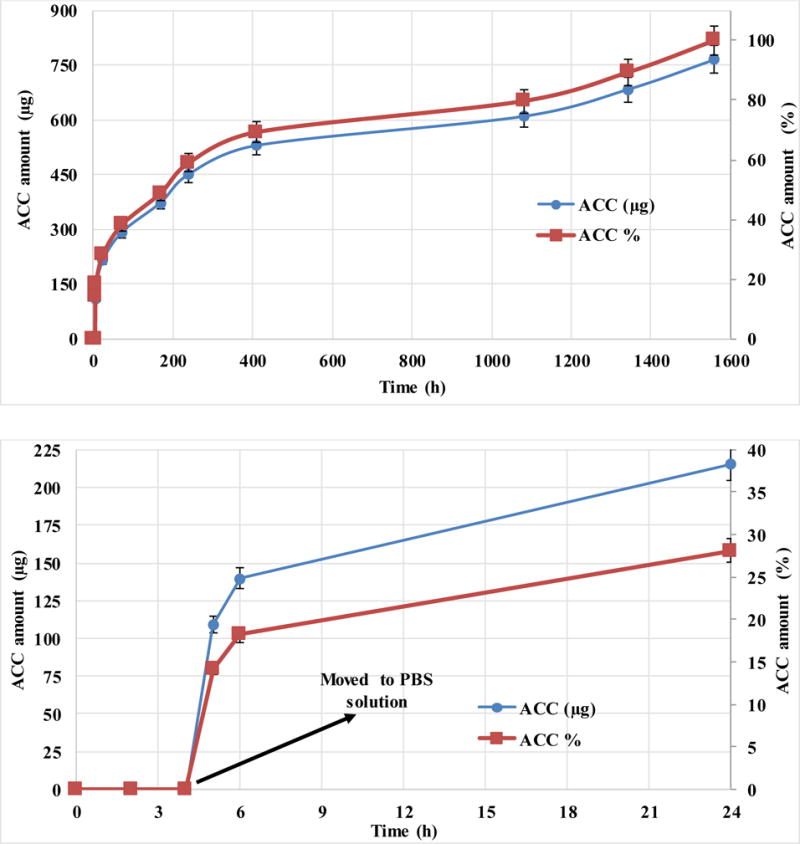
A quick fabrication method for making double-walled (DW) polymeric nanospheres is presented. The process uses sequential precipitation of two polymers. By choosing an appropriate solvent and non-solvent polymer pair, and engineering two sequential phase inversions which induces first precipitation of the core polymer followed by precipitation of the shell polymer, DW nanospheres can be created instantaneously. A series of DW formulations were prepared with various core and shell polymers, then characterized using laser diffraction particle sizing, scanning electron microscopy, atomic force microscopy, Fourier transform infrared spectroscopy, and differential scanning calorimetry (DSC). Atomic force microscopy (AFM) imaging confirmed existence of a single core polymer coated with a second polymer. Insulin (3.3% loading) was used as a model drug to assess its release profile from core (PLGA) and shell (PBMAD) polymers and resulted with a tri-phase release profile in vitro for two months. Current approaches for producing DW nanoparticles (NPs) are limited by the complexity and time involved. Additional issues include aggregation and entrapment of multiple spheres and the undesired formation of heterogeneous coatings. Therefore, the technique presented here is advantageous because it can produce NPs with distinct, core-shell morphologies through a rapid, spontaneous, self-assembly process. This method not only produces DW NPs, but can also be used to encapsulate therapeutic drug. Furthermore, modification of this process to other core and shell polymers is feasible using the general guidelines provided in this paper.

Keywords: AFM; DSC; Double-walled nanoparticles; Nanoparticles production method; Polymers' mixtures.


Figures

Figure 1
Figure 1
Top: Schematic of the Single Step Double Wall Nanoencapsulation (SSDN) process for the PLGA (75:25)/PBMAD DW formulation. Bottom (a, b, and c): Suggested mechanism of particle formation via the SSDN process; (a) cloud point of PLGA in THF-ethanol mixed solvent solution causing the PLGA to phase separate, while the PBMAD is fully soluble; (b) phase inversion of the PBMAD shell around the PLGA core in excess non-solvent for both polymers (petroleum ether); (c) DW PLGA-core/PBMAD-shell nanosphere created after the solvents (THF and ethanol) are leaching out.
Figure 2
Figure 2
Coulter laser particle size analysis (volume weighted) of nanospheres prepared by SSDN with different shell polymers and PMMA cores in aquatic solution.
Figure 3
Figure 3
Scanning electron micrographs of DW nanosphere formulations (a) PBMAD/PLGA (75:25), (b) PBMAD/PLGA (75:25), (c) PBMAD/PLGA (75:25), (d) PBMAD/PMMA 75:25, (e) PBMAD/PLGA (50:50), (f) PBMAD/D,L-PLA, 3000x, (g) PBMAP/PMMA 75:25, (h) PBMAT/PMMA 75:25, (i) PBMAD/PMMA 75:25, (j) PBMAP/PMMA 75:25, (k) PBMAT/PMMA 75:25, (l) PBMAP/PMMA 75:25; scale bars: 5 μm (a, d–l), 2.5 μm (b), 1.5 μm (c).
Figure 4
Figure 4
Fourier transform infrared spectroscopy (FTIR) analysis of PLGA (red), PBMAD (blue) and PBMAD-shell/PLGA-core (green) nanospheres; characteristic peaks have been identified and labeled.
Figure 5
Figure 5
AFM 3D images of NPs manufactured by the SSDN method (A) Pure PBMAD NPs (B) Pure PLGA 75:25 NPs, and (C) PBMAD/PLGA 75:25 DW NPs. XYZ axes represent scan size and NPs morphology while the purple-orange-yellow scale bar (same scale applied for all images) represent the phase angle shift data. Phase angle shift images were superimposed on top of the reciprocal 3D height images without any image color manipulations.
Figure 6
Figure 6
AFM 3D images of NPs of pure PLGA 75:25 NPs before (A) and after (B) exposure to pH=10 and PBMAD/PLGA 75:25 DW NPs before (C) and after (D) exposure to pH=10. XYZ axes provide length, width, and height scaling of the NPs while the purple-orange-yellow scale bar (same scale for all images) show the phase angle shift, which is indicative of material type. Phase angle shift images were superimposed on top of the reciprocal 3D height images without any image color manipulations.
Figure 7
Figure 7
Accumulated release profile of total insulin mass (μg, left Y-axis) and by percentage (%, right Y-axis) versus time of zinc bovine insulin from DW NPs of PLGA-core and PBMAD-shell. Theoretical loading was 5% (actual loading was 3.3%). The top figure shows the full-time scale of the experiment while the bottom image is just the first 24 hours of release (for better visualization of the profile). Values represent the average of three repeats with their respective standard deviations.

References

    1. Freiberg S, Zhu X. Polymer microspheres for controlled drug release. Int J Pharm. 2004;282:1–18. - PubMed
    1. Tan EC, Lin R, Wang C. Fabrication of double-walled microspheres for the sustained release of doxorubicin. J Colloid Interface Sci. 2005;291:135–143. - PubMed
    1. Pekarek KJ, Jacob JS, Mathiowitz E. Double-walled polymer microspheres for controlled drug release. Nature. 1994;367:258–260. - PubMed
    1. Xu Q, Xia Y, Wang C, Pack DW. Monodisperse double-walled microspheres loaded with chitosan-p53 nanoparticles and doxorubicin for combined gene therapy and chemotherapy. J Controlled Release. 2012;163:130–135. - PMC - PubMed
    1. Leach K. Effect of manufacturing conditions on the formation of double-walled polymer microspheres. J Microencapsul. 1999;16:153–167. - PubMed

Publication types