. 2018 Aug:68:68-75.
doi: 10.1016/j.neurobiolaging.2018.03.033. Epub 2018 Apr 5.

Interaction between a MAPT variant causing frontotemporal dementia and mutant APP affects axonal transport

Affiliations

Interaction between a MAPT variant causing frontotemporal dementia and mutant APP affects axonal transport

Robert Adalbert et al. Neurobiol Aging. 2018 Aug.

Abstract

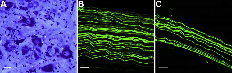
In Alzheimer's disease, many indicators point to a central role for poor axonal transport, but the potential for stimulating axonal transport to alleviate the disease remains largely untested. Previously, we reported enhanced anterograde axonal transport of mitochondria in 8- to 11-month-old MAPTP301L knockin mice, a genetic model of frontotemporal dementia with parkinsonism-17T. In this study, we further characterized the axonal transport of mitochondria in younger MAPTP301L mice crossed with the familial Alzheimer's disease model, TgCRND8, aiming to test whether boosting axonal transport in young TgCRND8 mice can alleviate axonal swelling. We successfully replicated the enhancement of anterograde axonal transport in young MAPTP301L/P301L knockin animals. Surprisingly, we found that in the presence of the amyloid precursor protein mutations, MAPTP301L/P3101L impaired anterograde axonal transport. The numbers of plaque-associated axonal swellings or amyloid plaques in TgCRND8 brains were unaltered. These findings suggest that amyloid-β promotes an action of mutant tau that impairs axonal transport. As amyloid-β levels increase with age even without amyloid precursor protein mutation, we suggest that this rise could contribute to age-related decline in frontotemporal dementia.

Keywords: Alzheimer's disease; Axonal transport; Aβ; FTDP-17T; Mitochondria; P301L mutation.


Figures

Fig. 1
Fig. 1
Changes in mitochondria transport in TgCRND8 mice homozygous for MAPTP301L tau knockin mutation. Quantification of anterograde (A and C) and retrograde (B and D) mitochondria transport in sciatic nerves from 3-month-old mice with indicated genotypes (all mice are MitoP positive). For all graphs, each data point represents the mean value obtained for 1 animal (5 fields of view and, on average, 15 axons per animal). Horizontal bar indicates mean and error bars indicate standard error of the mean. Statistically significant differences between genotypes are indicated (*p < 0.05, **p < 0.01; 1-way analysis of variance with Tukey multiple comparisons posttest). Abbreviation: n.s., not significant.
Fig. 2
Fig. 2
Human Aβ1–42 is present in peripheral nerves of TgCRND8 mice, and its levels in brain are independent of the MAPTP301L knockin mutation. (A) Concentration of human Aβ1–42 (ng/g) in sciatic nerves of 3-month-old wild-type (MitoP) and TgCRND8 mice. (B) Brain concentration of human Aβ1–42 (ng/g) in TgCRND8 and TgCRND8 mice heterozygotes for the MAPTP301L knockin mutation. Each data point represents 1 animal (mean and standard error of the mean, unpaired t test). Abbreviation: n.s., not significant.
Fig. 3
Fig. 3
Homozygous MAPTP301L knockin mutation does not reduce the number of plaques and the associated neurite swellings in TgCRND8 brains. (A) Amyloid plaques (arrows) in the cortex of 3-month-old TgCRND8 mouse and (B) a single amyloid plaque associated with neurite dystrophy (arrow). Quantification (mean and standard error of the mean, unpaired t test; n = 5) of the number of plaques (C), and plaques with associated dystrophy (D) reveals no significant difference (ns) between TgCRND8 and TgCRND8/MAPTP301L/P301L mice. Blue: Thioflavin S; Green: YFP in B. Scale bar: A, 100 μm; B, 20 μm. (For interpretation of the references to color in this figure legend, the reader is referred to the Web version of this article.)
Fig. 4
Fig. 4
Morphologically intact spinal cord motor neurons and peripheral nerves in 3-month-old TgCRND8/MAPTP301L/P301L mice. (A) Cresyl violet (Nissl)-stained transverse section through the L3-L5 lumbar spinal cord segment. (B) YFP positive axons from sciatic and (C) tibial nerves. Scale bar: A, 20 μm; B and C, 50 μm. (For interpretation of the references to color in this figure legend, the reader is referred to the Web version of this article.)
Fig. 5
Fig. 5
No tau histopathology in the brains of 6-month-old TgCRND8 mice homozygous for MAPTP301L tau knockin mutation. (A, B) PHF1 immunostaining in the cortex showed only puncta-like staining of dystrophic neurites (arrow) around the congo red positive plaques (star) with no neuronal staining in the mouse frontal cortex of TgCRND8 (A) or TgCRND8/MAPTP301L/P301L (B). (C–F) Gallyas silver staining. No NFTs was present in the mouse brains of TgCRND8 (C) or TgCRND8/MAPTP301L/P301L (D). Tg30 tau transgenic mouse brain expressing human P301S/G272V mutant tau (Leroy et al., 2007) (E) or human AD tissue (F) acting as positive control for NFT and dystrophic neurites around amyloid plaque (star). Scale bars: 20 μm. (For interpretation of the references to color in this figure legend, the reader is referred to the Web version of this article.)

References

    1. Adalbert R., Coleman M.P. Review: axon pathology in age-related neurodegenerative disorders. Neuropathol. Appl. Neurobiol. 2013;39:90–108. - PubMed
    1. Adalbert R., Nogradi A., Babetto E., Janeckova L., Walker S.A., Kerschensteiner M., Misgeld T., Coleman M.P. Severely dystrophic axons at amyloid plaques remain continuous and connected to viable cell bodies. Brain. 2009;132(Pt 2):402–416. - PubMed
    1. Adalbert R., Nogradi A., Szabo A., Coleman M.P. The slow Wallerian degeneration gene in vivo protects motor axons but not their cell bodies after avulsion and neonatal axotomy. Eur. J. Neurosci. 2006;24:2163–2168. - PubMed
    1. Adams S.J., Crook R.J., Deture M., Randle S.J., Innes A.E., Yu X.Z., Lin W.L., Dugger B.N., McBride M., Hutton M., Dickson D.W., McGowan E. Overexpression of wild-type murine tau results in progressive tauopathy and neurodegeneration. Am. J. Pathol. 2009;175:1598–1609. - PMC - PubMed
    1. Allen B., Ingram E., Takao M., Smith M.J., Jakes R., Virdee K., Yoshida H., Holzer M., Craxton M., Emson P.C., Atzori C., Migheli A., Crowther R.A., Ghetti B., Spillantini M.G., Goedert M. Abundant tau filaments and nonapoptotic neurodegeneration in transgenic mice expressing human P301S tau protein. J. Neurosci. 2002;22:9340–9351. - PMC - PubMed

Publication types

MeSH terms