. 2018 Jul 15;368(2):225-235.
doi: 10.1016/j.yexcr.2018.05.001. Epub 2018 May 4.

iPSC-derived neurons profiling reveals GABAergic circuit disruption and acetylated α-tubulin defect which improves after iHDAC6 treatment in Rett syndrome

Affiliations

iPSC-derived neurons profiling reveals GABAergic circuit disruption and acetylated α-tubulin defect which improves after iHDAC6 treatment in Rett syndrome

Elisa Landucci et al. Exp Cell Res. .

Abstract

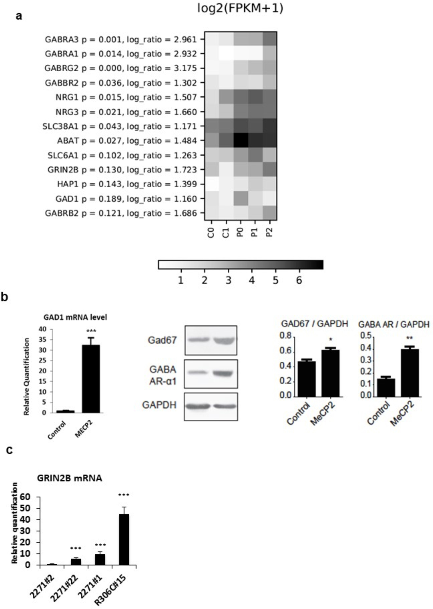
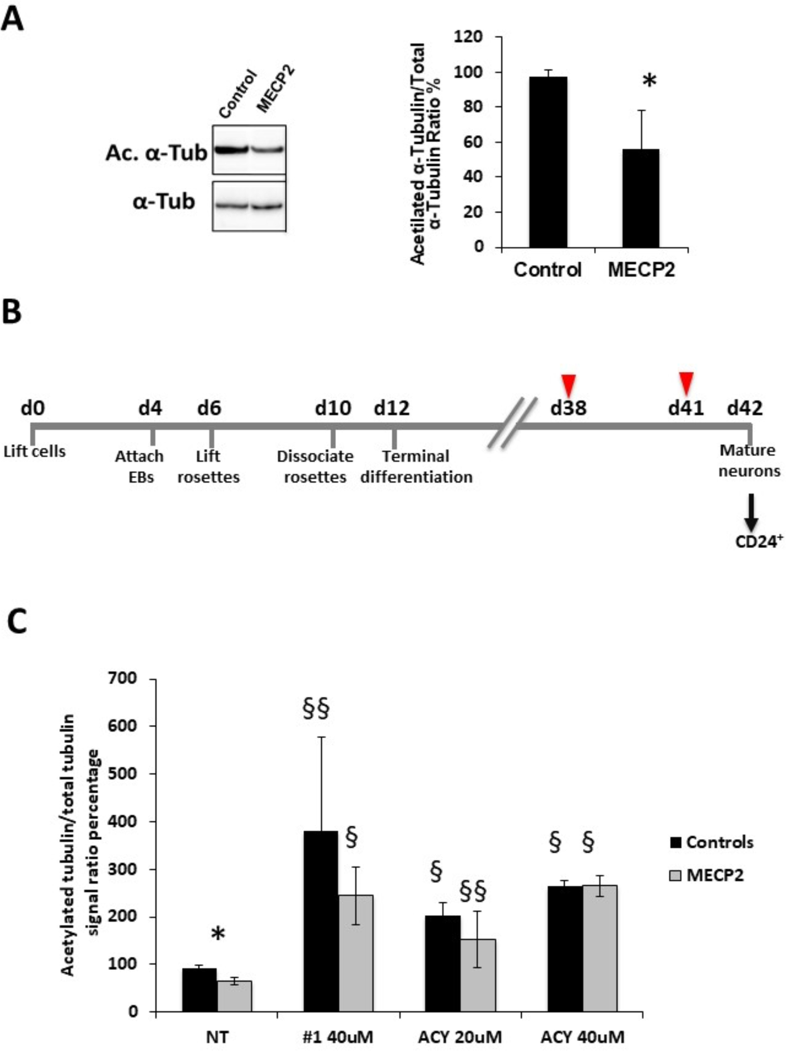
Mutations in MECP2 gene have been identified in more than 95% of patients with classic Rett syndrome, one of the most common neurodevelopmental disorders in females. Taking advantage of the breakthrough technology of genetic reprogramming, we investigated transcriptome changes in neurons differentiated from induced Pluripotent Stem Cells (iPSCs) derived from patients with different mutations. Profiling by RNA-seq in terminally differentiated neurons revealed a prominent GABAergic circuit disruption along with a perturbation of cytoskeleton dynamics. In particular, in mutated neurons we identified a significant decrease of acetylated α-tubulin which can be reverted by treatment with selective inhibitors of HDAC6, the main α-tubulin deacetylase. These findings contribute to shed light on Rett pathogenic mechanisms and provide hints for the treatment of Rett-associated epileptic behavior as well as for the definition of new therapeutic strategies for Rett syndrome.

Keywords: GABA; HDAC6; HDAC6 inhibitors; RNA-seq; acetylated α-tubulin; iPSC‐derived neurons.


Conflict of interest statement

Declaration of interest

Conflicts of interest: none.

Figures

Fig. 1
Fig. 1. RNAseq data analysis.
a) Scatter plot of log2 transformed RNAseq expression level (FPKM) of MECP2-derived cells (P0, P1, P2) versus controls (C0, C1) in terminally differentiated neurons (day 30 is) shown. Red and green spots indicate significantly (p<0.05) up-regulated and down-regulated genes, respectively.
Fig. 2
Fig. 2. Up regulation of GABAergic axis.
a) HeatMap of GABAergic pathway genes; for each gene, log ratio and related p-values are shown. b) Validation of RNAseq data on neurons from and independent differentiation experiment demonstrate a significant upregulation of GAD1 mRNA and protein levels and of GABA-R receptor alpha 1 (GABA-AR) protein in MECP2-mutated neurons compared to controls. GAPDH was used as loading control for western blot analysis. n=3. c) Upregulation of GRIN2B mRNA in neurons from the 3 MECP2-mutated clones respect to neurons from the partial isogenic control clone (2271#2). Statistical significance was determined using unpaired student’s t test (*p<0.05; **p<0.001; ***p<0.0001).
Fig. 3
Fig. 3. Altered functional properties of MeCP2 mutant neurons.
a) Membrane resting potential (mV). Student t test: p = 0.093, n wt = 32, n ko = 22. b) Membrance capacitance (pF), p = 0.063, n wt = 48, n ko = 24. c) Membrane resistance (MΩ). Student t test: p = 0.31, n wt = 43, n ko = 24. d) Maximum peak of Na current recorded in voltage clamp. Student t test: p = 0.373, n wt = 33, n ko = 23. e) Firing: number of action potentials evoked by +10 mV steps of the membrane potential starting from −70 mV. Two way ANOVA, * p < 0.05, n wt = 27, n ko = 20. f) Example of traces for control and MECP2 mutated cells; traces from the same cell in the presence of bicuculine are shown on the right. g) Average sIPSCs in a control (CNT) and a mutant cell. h) Cumulative distribution of sIPSC amplitude (pA). Mann-Whitney: p <0.001; wt median: 47.0, 25th-75th percentile = 30.0–87.0; ko median: 40.1, 25th-75th percentile = 24.5– 73.0. i) Cumulative distribution of sIPSCs inter-event interval (ms). Mann-Whitney: p <0.001; wt median: 56.4, 25th-75th percentile: 263.5–1494.6; ko median: 895.0, 25th-75th percentile 392.6–1764.0. j) Whisker plot of sIPSC decay kinetics (90–10% decay time). Mann-Whitney: p = 0.078; wt: median: 28.6, 25th-75th percentile 18.6–37.7; ko median: 25.3, 25th-75th percentile 15.2–41.8. Measurements for h, i and j were collected from 28 control neurons and 18 MECP2-mutated neurons. Data are reported as mean ± SEM. Each circle in a-d represents a cell.
Fig. 4.
Fig. 4.. Reduction in acetylated α-tubulin levels and treatment with selective HDAC6 inhibitors.
a) A reduction in acetylated α-tubulin levels was observed by Western Blot analysis in MECP2-derived neurons (n=3 clones) compared to control ones (n=2 clones). * p<0.05. b) Overview of the neuronal differentiation protocol with critical time-points indicated below the time lane. Cells were differentiated in Terminal Differentiation medium for 30 days; differentiating cells were treated twice (arrowheads) with either the selective inhibitor (Compound #1 or ACY1215) or vehicle and then neurons were isolated (arrow) by immunomagnetic sorting with anti-CD24 antibodies for further analyses. c) Treatment with compound #1 (40uM) or ACY-1215 (ACY 20uM or ACY 40uM) resulted in a significant increase in acetylated α-tubulin levels in both MECP2-mutated and control neurons. Data are expressed as the mean ± SEM from 3 independent experiments performed on 3 control and 3 MECP2-mutated clones. *p<0.05 between mutated and control neurons; §p<0.0001 and §§p<0.005 compared to the corresponding untreated neurons. Statistical significance was determined using unpaired student’s t test.

References

    1. Chahrour M, Zoghbi HY, The story of Rett syndrome: from clinic to neurobiology, Neuron 56 (2007) 422–437. - PubMed
    1. Katz DM, Bird A, Coenraads M, Gray SJ, Menon DU, Philpot BD, Tarquinio DC, Rett Syndrome: Crossing the Threshold to Clinical Translation, Trends Neurosci 39 (2016) 100–113. - PMC - PubMed
    1. Hagberg B, Witt-Engerstrom I, Rett Syndrome: A suggested staging system for describing impairment profile with increasing age towards adolescence, Am J Hum Genet 24 (1986) 47–59. - PubMed
    1. Grillo E, Villard L, Clarke A, Ben Zeev B, Pineda M, Bahi-Buisson N, Hryniewiecka-Jaworska A, Bienvenu T, Armstrong J, Roche-Martinez A, Mari F, Veneselli E, Russo S, Vignoli A, Pini G, Djuric M, Bisgaard AM, Mejaski Bosnjak V, Polgar N, Cogliati F, Ravn K, Pintaudi M, Melegh B, Craiu D, Djukic A, Renieri A, Rett networked database: an integrated clinical and genetic network of Rett syndrome databases, Hum Mutat 33 (2012) 1031–1036. - PubMed
    1. Lyst MJ, Bird A, Rett syndrome: a complex disorder with simple roots, Nat Rev Genet 16 (2015) 261–275. - PubMed

Publication types