Review
. 2018 May:29:207-214.
doi: 10.1016/j.scr.2018.04.009. Epub 2018 Apr 21.

Spermatogonial stem cells and spermatogenesis in mice, monkeys and men

Affiliations
Review

Spermatogonial stem cells and spermatogenesis in mice, monkeys and men

Adetunji P Fayomi et al. Stem Cell Res. 2018 May.

Abstract

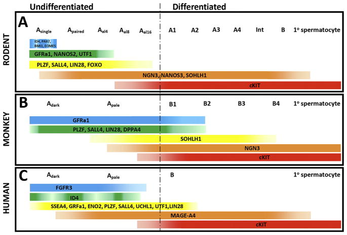
Continuous spermatogenesis in post-pubertal mammals is dependent on spermatogonial stem cells (SSCs), which balance self-renewing divisions that maintain stem cell pool with differentiating divisions that sustain continuous sperm production. Rodent stem and progenitor spermatogonia are described by their clonal arrangement in the seminiferous epithelium (e.g., Asingle, Apaired or Aaligned spermatogonia), molecular markers (e.g., ID4, GFRA1, PLZF, SALL4 and others) and most importantly by their biological potential to produce and maintain spermatogenesis when transplanted into recipient testes. In contrast, stem cells in the testes of higher primates (nonhuman and human) are defined by description of their nuclear morphology and staining with hematoxylin as Adark and Apale spermatogonia. There is limited information about how dark and pale descriptions of nuclear morphology in higher primates correspond with clone size, molecular markers or transplant potential. Do the apparent differences in stem cells and spermatogenic lineage development between rodents and primates represent true biological differences or simply differences in the volume of research and the vocabulary that has developed over the past half century? This review will provide an overview of stem, progenitor and differentiating spermatogonia that support spermatogenesis; identifying parallels between rodents and primates where they exist as well as features unique to higher primates.

Keywords: A(dark); A(pale); Male fertility; Spermatogenic lineage development; Spermatogonial stem cells; Stem cells; Testis.


Figures

Fig. 1
Fig. 1
Clonal development in the spermatogenic lineages of rodents, monkeys and humans. Undifferentiated spermatogonia are described as As, Apr or Aal in the rodents and Adark or Apale in monkey and human. During spermatogenic development, Asingle (As) and Adark and/or Apale undergo one or more mitotic divisions to give rise to cells of larger clones (chains) of interconnected cells sizes through transit-amplifying mitotic divisions. A) Clonal development in rodents features 3–4 transit amplifying divisions in the pool of undifferentiated As, Apr and Aal spermatogonia followed by 6 amplifying divisions in the pool of differentiated spermatogonia (A1–A4, Intermediate, B), which give rise to primary spermatocytes. Two additional meiotic divisions produce round spermatids that undergo spermiogenesis to produce sperm. B) Clonal development of spermatogonia in monkeys features 0, 1 or 2 transit amplifying divisions in the pool of undifferentiated Adark/Apale spermatogonia, followed by 4 transit amplifying divisions of differented spermatogonia (B1–B4), which give rise to primare spermatocytes. C) Clonal development of spermatogonia in humans features 0, 1 or 2 transit amplifying divisions in the pool of undifferentiated Adark/Apale spermatogonia followed by a single a single transit amplyfying division in differentiated B spermatogonia that give rise to primary spermatocytes. Thus, there are typically 12 transit amplifying divisions in rodents; 8 in monkeys and 5 in humans between stem cell and sperm. The reduced number of transit amplyfing divisions in monkeys and humans is compensated in part by a larger stem cell pool (see Fig. 2).
Fig. 2
Fig. 2
Schematic comparison of the stem cell pools and sperm output in mice, monkeys and humans. A) The spermatogenic lineage in mice features a relatively small pool of As, Apr and Aal undifferentiated spermatogonia (0.3% of germ cells). However, with about 12 transit amplifying divisions between stem cells and sperm (see Fig. 1), the small pool of stem cells produced 40 million sperm per gram of testicular tissue per day. B) The spermatogenic lineage in monkeys features a relatively larger pool of Adark/Apale spermatogonia (4% of germ cells) and this compensates for the reduced number of transit amplying divisions between stem cell and sperm. Sperm output in monkeys is similar to mice: 41 million sperm per gram of testicular tissue per day. C) The spermatogenic lineage in humans features the largest pool of undifferentiated Adark/Apale spermatogonia (22% of germ cells). However, with only one transit amplifying division of differentiated spermatogonia, sperm output in humans is reduced to 4.4 million sperm per gram of testicular tissue per day. Thus distinguishing features of spermatogenic lineage development in mice, monkeys and men include 1) the size of the pool of undifferentiated stem/progenitor spermatogonia; 2) the number of transit amplifying divisions in differentiated spermatogonia and 3) sperm output.
Fig. 3
Fig. 3
Spermatogonial markers in rodents, monkeys and humans. A) Rodents; B) Monkeys; C) Humans. Several markers are conserved from rodents to monkeys to humans, suggesting their importance in spermatogenic lineage development. GFRa1, PLZF, SALL4 and LIN28 are conserved markers of undifferentiated spermatogonia. cKIT is a conserved marker of differentiating/differentiated spermatogonia. The following references describe markers in this figure that were not referenced elsewhere in the text: NGN3 (Yoshida et al., 2004); SOHLH1 (Ballow et al., 2006).
Fig. 4
Fig. 4
Histological and immunohistochemical evaluation of Adark and Apale spermatogonia in monkeys and humans. Periodic acid, Schiffs’ and Hematoxylin (PAS-H) staining in monkey (A and C) and human (B and D) testis section reveals Adark (black arrows) and Apale (red arrows) spermatogonia on the basement membrane of the seminiferous tubules. The sub-population of Adark spermatogonia with a rarefraction zone are indicated by a green arrow in (B). Colorimetric staining for UTF1 (brown color) with PAS-H staining confirms that UTF1 is a conserved marker of most, but not all Adark (black arrow) and Apale (red arrows) spermatogonia in monkey (C) and Human (D) testes. (For interpretation of the references to color in this figure legend, the reader is referred to the web version of this article.)

References

    1. Aeckerle N, Eildermann K, Drummer C, Ehmcke J, Schweyer S, Lerchl A, Bergmann M, Kliesch S, Gromoll J, Schlatt S, Behr R. The pluripotency factor LIN28 in monkey and human testes: a marker for spermatogonial stem cells? Mol Hum Reprod. 2012;18:477–488. - PMC - PubMed
    1. Aloisio GM, Nakada Y, Saatcioglu HD, Pena CG, Baker MD, Tarnawa ED, Mukherjee J, Manjunath H, Bugde A, Sengupta AL, Amatruda JF, Cuevas I, Hamra FK, Castrillon DH. PAX7 expression defines germline stem cells in the adult testis. J Clin Invest. 2014;124:3929–3944. - PMC - PubMed
    1. Ballow D, Meistrich ML, Matzuk M, Rajkovic A. Sohlh1 is essential for spermatogonial differentiation. Dev Biol. 2006;294:161–167. - PubMed
    1. Braun RE, Sharma M, Srivastave A, Fairfield HE, Bergstrom DE. Identification of Slow-Cycling Long-Term Spermatogonial Stem Cells and their Regulation by PLZF. Society for the Study of Reproduction; Washington DC: 2017. p. 279.
    1. Brinster RL, Avarbock MR. Germline transmission of donor haplotype following spermatogonial transplantation. Proc Natl Acad Sci U S A. 1994;91:11303–11307. - PMC - PubMed

Publication types