Review
. 2018 Jun;11(2):298-306.
doi: 10.1007/s12178-018-9488-x.

The Role of Osteotomy for the Treatment of PCL Injuries

Affiliations
Review

The Role of Osteotomy for the Treatment of PCL Injuries

João V Novaretti et al. Curr Rev Musculoskelet Med. 2018 Jun.

Abstract

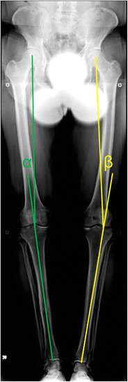
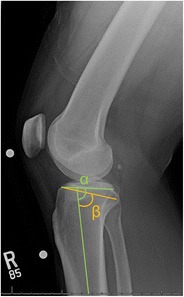
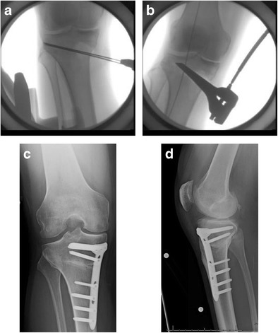
Purpose of review: The purposes of this review are to (1) describe the anatomic and biomechanical rationale for high tibial osteotomy (HTO) in the setting of posterior cruciate ligament (PCL) deficiency, (2) review the indications for concomitant HTO and PCL reconstruction, (3) provide guidance for the clinical assessment of the patient with suspected PCL deficiency, and (4) summarize the key surgical steps necessary to attain the appropriate sagittal and coronal plane corrections.

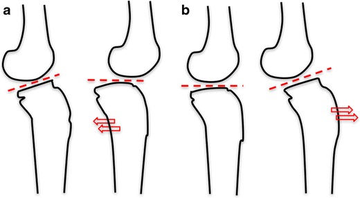
Recent findings: The preponderance of available biomechanical data pertaining to the PCL-deficient knee suggests that an increased proximal tibial slope limits posterior tibial translation under axial compressive loads. Moreover, recent clinical data has demonstrated that decreased proximal tibial slope may exacerbate residual anterior-posterior laxity and jeopardize the durability of PCL reconstruction. Thus, in the setting of PCL deficiency, an HTO that increases the posterior tibial slope may be advisable. HTO may be an important treatment adjunct in the surgical management of PCL deficiency. In the setting of chronic injuries and varus malalignment, HTO should be considered in order to ensure a durable ligamentous reconstruction and forestall the progression of secondary osteoarthritis.

Keywords: High tibial osteotomy (HTO); Posterior cruciate ligament (PCL); Posterior tibial translation; Tibial slope; Varus malalignment.


Conflict of interest statement

Conflict of Interest

All authors declare that they have no conflict of interest.

Human and Animal Rights and Informed Consent

This article does not contain any studies with human or animal subjects performed by any of the authors.

Figures

Fig. 1
Fig. 1
Single cassette, full-length anterior posterior standing radiograph of bilateral lower extremities is used to calculate required correction of varus malalignment in the coronal plane. Right knee: When OA changes are not present in the medial compartment, the correction should target for the mechanical axis to cross the tibial plateau at 50% of total tibial plateau width. The angle α represents the required correction. Left knee: If OA in the medial compartment is present, the correction should target for the mechanical axis to cross the tibial plateau at 62% of total tibial plateau width, from medial to lateral. The angle β represents the required correction
Fig. 2
Fig. 2
Four methods for measuring the native tibial slope using lateral radiographs of the knee. a Anterior tibial cortex (ATC) [60]. b Proximal tibial anatomic axis (PTAA) [51]. c Posterior tibial cortex (PTC). d Proximal fibular anatomic axis (PFA) [48]
Fig. 3
Fig. 3
Changes in the tibial slope and consequences in the anteroposterior tibial translation. a A decrease in the tibial slope leads to a posterior translation of tibia, stabilizing knees with ACL deficiency. b An increase in the tibial slope promotes anterior translation of the tibia, improving stability of PCL-deficient knees
Fig. 4
Fig. 4
Preoperative plan for sagittal correction in PCL deficiency. Green: lines for measurement of tibial slope using the proximal tibial anatomic axis (PTAA) method; α angle: decreased tibial slope angle in a PCL-deficient knee; β angle: desired increase in tibial slope angle
Fig. 5
Fig. 5
Medial opening wedge HTO. a Introduction of two kirschner wires aimed toward the proximal tibiofibular joint (intraoperative fluoroscopic image). b Laminar spreader is introduced anteriorly and the desired opening from preoperative measurements is achieved to simultaneously correct varus and increase tibial slope if so desired (intraoperative fluoroscopic image). c, d Locking plate application to the medial proximal tibia cortex to maintain desired correction and stabilize the osteotomy (postoperative radiographs)

References

    1. Butler DL, Noyes FR, Grood ES. Ligamentous restraints to anterior-posterior drawer in the human knee. A biomechanical study. J Bone Joint Surg Am. 1980;62(2):259–270. doi: 10.2106/00004623-198062020-00013. - DOI - PubMed
    1. Fukubayashi T, Torzilli PA, Sherman MF, Warren RF. An in vitro biomechanical evaluation of anterior-posterior motion of the knee. Tibial displacement, rotation, and torque. J Bone Joint Surg Am. 1982;64(2):258–264. doi: 10.2106/00004623-198264020-00018. - DOI - PubMed
    1. Girgis FG, Marshall JL, Monajem A. The cruciate ligaments of the knee joint. Anatomical, functional and experimental analysis. Clin Orthop Relat Res. 1975;106:216–231. doi: 10.1097/00003086-197501000-00033. - DOI - PubMed
    1. Gollehon DL, Torzilli PA, Warren RF. The role of the posterolateral and cruciate ligaments in the stability of the human knee. A biomechanical study. J Bone Joint Surg Am. 1987;69(2):233–242. doi: 10.2106/00004623-198769020-00010. - DOI - PubMed
    1. Grood ES, Stowers SF, Noyes FR. Limits of movement in the human knee. Effect of sectioning the posterior cruciate ligament and posterolateral structures. J Bone Joint Surg Am. 1988;70(1):88–97. doi: 10.2106/00004623-198870010-00014. - DOI - PubMed

LinkOut - more resources