. 2018 Jun 14;173(7):1783-1795.e14.
doi: 10.1016/j.cell.2018.03.061. Epub 2018 May 3.

Identification of Near-Pan-neutralizing Antibodies against HIV-1 by Deconvolution of Plasma Humoral Responses

Affiliations

Identification of Near-Pan-neutralizing Antibodies against HIV-1 by Deconvolution of Plasma Humoral Responses

Mohammad Mohseni Sajadi et al. Cell. .

Abstract

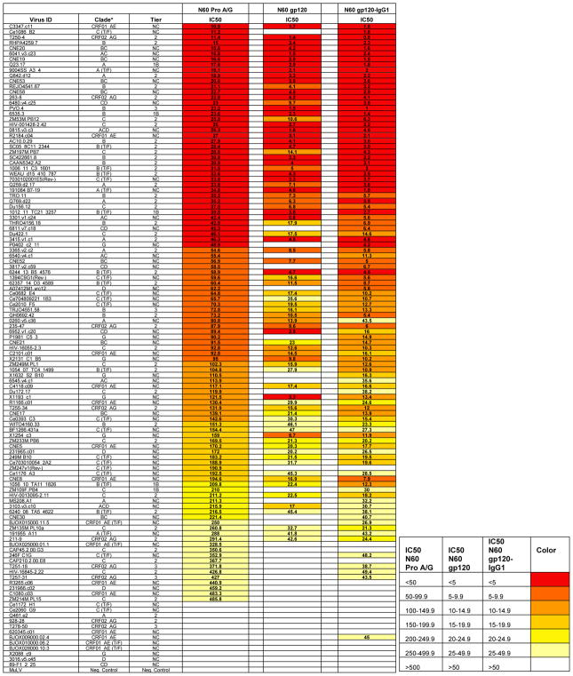
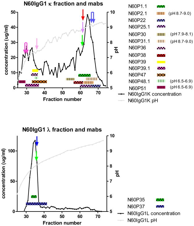
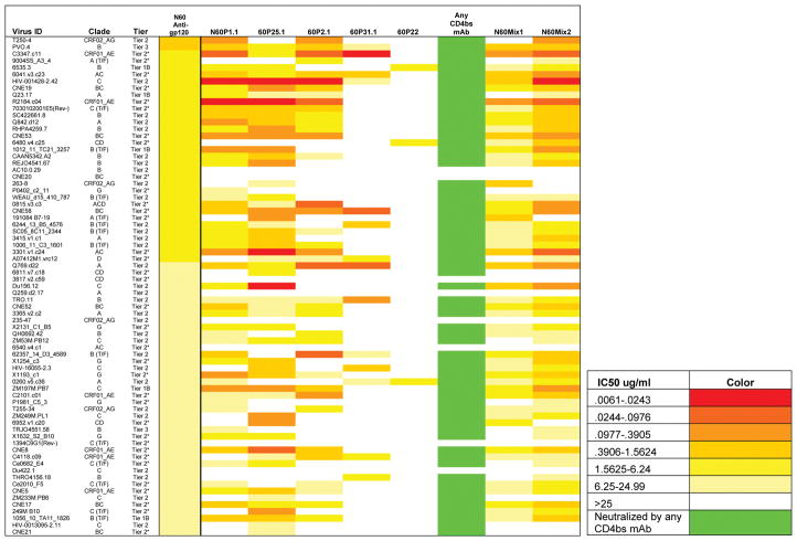
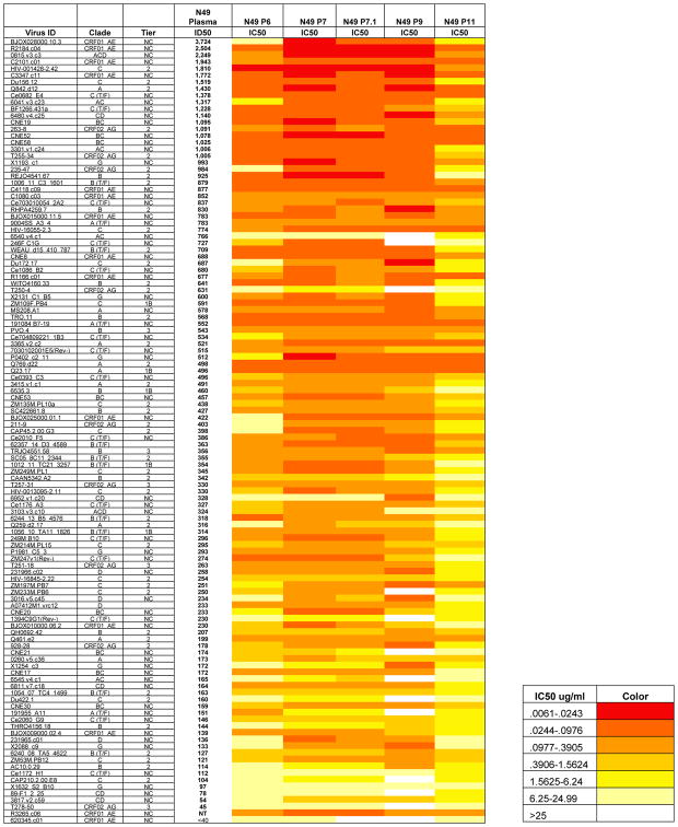
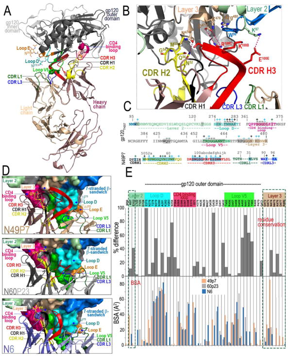
Anti-HIV-1 envelope broadly neutralizing monoclonal antibodies (bNAbs) isolated from memory B cells may not fully represent HIV-1-neutralizing profiles measured in plasma. Accordingly, we characterized near-pan-neutralizing antibodies extracted directly from the plasma of two "elite neutralizers." Circulating anti-gp120 polyclonal antibodies were deconvoluted using proteomics to guide lineage analysis of bone marrow plasma cells. In both subjects, a single lineage of anti-CD4-binding site (CD4bs) antibodies explained the plasma-neutralizing activity. Importantly, members of these lineages potently neutralized 89%-100% of a multi-tier 117 pseudovirus panel, closely matching the specificity and breadth of the circulating antibodies. X-ray crystallographic analysis of one monoclonal, N49P7, suggested a unique ability to bypass the CD4bs Phe43 cavity, while reaching deep into highly conserved residues of Layer 3 of the gp120 inner domain, likely explaining its extreme potency and breadth. Further direct analyses of plasma anti-HIV-1 bNAbs should provide new insights for developing antibody-based antiviral agents and vaccines.

Keywords: HIV-1; antibody; broadly neutralizing antibody; humoral immunity; pan-neutralization; repertoire.


Conflict of interest statement

Declaration of Interests

The authors declare no competing interests.

Figures

Figure 1
Figure 1. Neutralization of N60 parent and affinity purified samples
Pro A/G= Affinity purified antibody from a Protein A/G column; gp120= Affinity purified antibody from a monomeric gp120 column; gp120-IgG1= Affinity purified antibody from a gp120 followed by IgG1 column; IC50= Inhibitory concentration 50 (ug/ml); T/F= transmitted/founder; NC= not characterized. Total IgG from patient N60 was purified by Protein A/G and tested against a 118 multitier and multiclade pseudovirus panel (starting titer 528ug/ml). Parent sample demonstrates considerable breadth, which was also seen in the gp120 and gp120-IgG1 (starting titer 50ug/ml).
Figure 2
Figure 2. Free flow electrophoretic fractionation of N60 plasma anti-gp120 polyclonal antibodies and reconstructed anti-gp120 mAbs
The gray line indicates the pH (right Y axis) gradient across the fractions created by the FFE procedure. Anti-gp120 κ light chain (top) or anti-gp120 λ light chain (bottom) polyclonal plasma antibody preparations were processed separately (see main text and Methods). The plasma antibody protein concentrations (left Y axis) detected across fractions are shown by the black trace. FFE analyses of identified and reconstructed mAbs (see main text) are depicted by horizontal bars. Bar width spans 75–85% of the total amount of antibodies in the FFE fraction, as determined by ELISA. Eight mAbs (checkered bars) were identified by evaluating peptides from individual FFE fractions of anti-gp120 plasma antibodies. The FFE fraction reflecting the most coverage and unique peptide pairings is indicted for each mAb by a matched-color arrow. Four additional mAbs (hatched colored bars) were identified by evaluating peptides from IEF gel fractionation. Searches using peptides digested from bulk plasma anti-gp120 antibodies identified 1 additional mAbs (solid colored bars). One other mAb (criss-cross colored bars) was identified by homology search of the Ig gene database. The pH gradient shown is for the polyclonal N60IgG1 anto-gp120 κ fraction; pH gradients from each monoclonal run overlapped the trace shown, with a variance of up to 0–5 fractions in either direction.
Figure 3
Figure 3. Neutralization activity of N60 plasma derived anti-Env antibodies (alone and in combination)
A panel of pseudoviruses representing the indicated HIV-1 strains (listed in the left column) that were sensitive to the bulk plasma N60 gp120-Ig were tested against neutralizing anti-CD4bs antibodies from Lineage 1 and 2. For Lineage 2, only one mAb N60P22 was tested, as the other was a closely related clone (98% sequence homology). IC50 values are color-coded according to the color key on the left: the greater the neutralization, the darker red the color; grey represents no neutralization (IC50>25ug/ml). Taken together, the anti-CD4bs mAbs neutralized 89% of the viruses that were sensitive to bulk plasma anti-gp120 Ig. An equimolar mix of the mAbs called N60mAb Mix1 (all CD4bs, CD4i, and variable loop antibodies with > 5% sequence divergence) was tested at equimolar concentrations neutralized 79% of the pseudoviruses, and N60mAb Mix2 (all CD4bs antibodies with > 5% sequence divergence at equimolar concentrations) neutralized 89% of the pseudoviruses. IC50=Inhibitory Concentration 50 in ug/ml.
Figure 4
Figure 4. Neutralization activity of N49 plasma and P series mAbs
A “global panel” of 117 HIV-1 psuedovirus Tier 1-3 isolates (individual viruses listed on the left column) were tested against all N49 plasma and CD4bs antibodies. IC50 values are color-coded according to the color key on the left: the greater the neutralization, the darker red the color; white represents no neutralization (IC50>50ug/ml). The individual mAbs showed extreme breadth with N49P6, N49P7, and N49P11 exhibiting 100% breadth, N49P7.1 exhibiting 99% breadth, and N49P9 exhibiting 89% breadth. IC50=Inhibitory Concentration 50 in ug/ml. NC=not classified
Figure 5
Figure 5. Comparison of potency and breadth of resistant viruses between N49P7, N6, DH511-2, and 10E8
Viruses resistant to N6, DH511-2, or 10E8 are shown (IC50 values for DH511-2 obtained from (Williams et al., 2017)). N49P7 shows the greatest breadth and overall potency. NT = Not tested
Figure 6
Figure 6. Crystal structure of N49P7 Fab-gp12093TH057 coree complex
(A) Ribbon diagram of complex with the complementarity-determining regions (CDRs) of N49P7 Fab contributing to the gp120 binding (from light chain CDR L1 and CDR L3 and from heavy chain CDR H1, CDR H2 and CDR H3, see also Table S6) colored as shown. The gp120 outer and inner domains are colored in black and gray, respectively. The D (S274 – T283), E5 (F353 – T358), V5 (T455 - N465) and the CD4 binding (Q362 - G372) loops are colored in cyan, orange, violet and magenta, respectively. G54 of N49P7 Fab is highlighted in red and sugars at positions 276 (Loop D) and 355 (Loop E) are shown as sticks. (B) A blow up view into the network of interactions of N49P7 Fab with residues of the inner domain of gp120. Inner domain Layers 2 and 3 are colored pale green and beige, respectively. Two hydrogen bonds (CDR H2 N52 and Layer 3 N474; CDR H3 E100E and Layer 2 K97) and a salt bridge (CDR H3 R100C and Layer 3 D477) are formed at the N49P7 Fab – gp120 inner domain interface. (C) Gp12093TH057 coree and N49P7 CDR contact residues mapped onto the primary sequence. Contact residues are defined by a 5 Å cutoff and marked above the sequence. Side chain (+) and main chain (−) contacts are colored based on contact type; hydrophobic in green, hydrophilic in blue, or both in black. Buried surface residues as determined by PISA are shaded. (D) Blow up views into the Fab - gp12093TH057 coree interfaces of N49P7 Fab, N60P23 Fab and N6 Fab (PDB code: 5te6). The molecular surface is displayed over gp120 molecules with outer domain loops: D, X5, V5 and CD4 binding and inner domain Layers 2 and 3 and the 7-stranded β-sandwich with CDRs colored as in panels A and B. The Fab residue at the position equivalent to F43 of CD4 is shown as sticks (Gly, His and Tyr in N49P7, N60P23 and N6, respectively). (E) The buried surface area (BSA) of gp120 residues involved in binding to N49P7, N60P23 and N6 are shown as bars (bottom panel) and their conservation among HIV-1 isolates (top panel). The conservation of the residue at particular position is shown as % difference from the HXBc2 sequence. Only unique sequences in the database having an equivalent residue at each position were included in the calculated percentage representing approximately 32,000 sequences on average. Conserved inner domain residues uniquely targeted by N49P7 are highlighted in red.

References

    1. Boutz DR, Horton AP, Wine Y, Lavinder JJ, Georgiou G, Marcotte EM. Proteomic identification of monoclonal antibodies from serum. Analytical chemistry. 2014;86:4758–4766. - PMC - PubMed
    1. Briney BS, Willis JR, Finn JA, McKinney BA, Crowe JE., Jr Tissue-specific expressed antibody variable gene repertoires. PLoS One. 2014;9:e100839. - PMC - PubMed
    1. Brunger AT. Free R value: Cross-validation in crystallography. Method Enzymol. 1997;277:366–396. - PubMed
    1. de Taeye SW, Ozorowski G, Torrents de la Pena A, Guttman M, Julien JP, van den Kerkhof TL, Burger JA, Pritchard LK, Pugach P, Yasmeen A, et al. Immunogenicity of Stabilized HIV-1 Envelope Trimers with Reduced Exposure of Non-neutralizing Epitopes. Cell. 2015;163:1702–1715. - PMC - PubMed
    1. Dey B, Berger EA. Blocking HIV-1 gp120 at the Phe43 cavity: if the extension fits. Structure. 2013;21:871–872. - PMC - PubMed

Publication types

MeSH terms