

Pathophysiology of keratinization
- PMID: 29731562
- PMCID: PMC5917548
- DOI: 10.4103/jomfp.JOMFP_195_16
Pathophysiology of keratinization
Abstract
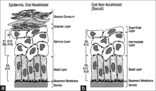
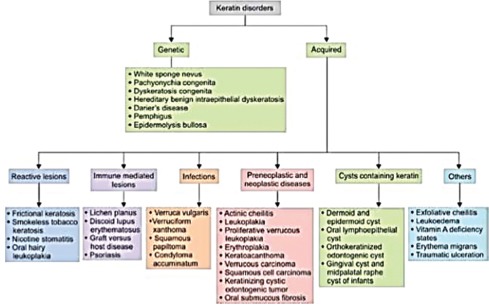
Cytoskeleton of a cell is made up of microfilaments, microtubules and intermediate filaments. Keratins are diverse proteins. These intermediate filaments maintain the structural integrity of the keratinocytes. The word keratin covers these intermediate filament-forming proteins within the keratinocytes. They are expressed in a specific pattern and according to the stage of cellular differentiation. They always occur in pairs. Mutations in the genes which regulate the expression of keratin proteins are associated with a number of disorders which show defects in both skin and mucosa. In addition, there are a number of disorders which are seen because of abnormal keratinization. These keratins and keratin-associated proteins have become important markers in diagnostic pathology. This review article discusses the classification, structure, functions, the stains used for the demonstration of keratin and associated pathology. The review describes the physiology of keratinization, pathology behind abnormal keratin formation and various keratin disorders.
Keywords: Keratinization; disorder; marker.
Conflict of interest statement
There are no conflicts of interest.
Figures
References
-
- Rao RS, Patil S, Ganavi BS. Oral cytokeratins in health and disease. J Contemp Dent Pract. 2014;15:127–36. - PubMed
-
- Vaidya MM, Kanojia D. Keratins: Markers of cell differentiation or regulators of cell differentiation? J Biosci. 2007;32:629–34. - PubMed
-
- Gu LH, Coulombe PA. Keratin function in skin epithelia: A broadening palette with surprising shades. Curr Opin Cell Biol. 2007;19:13–23. - PubMed
