. 2018 Apr 26:10:873-885.
doi: 10.2147/CMAR.S160172. eCollection 2018.

Liver-enriched activator protein 1 as an isoform of CCAAT/enhancer-binding protein beta suppresses stem cell features of hepatocellular carcinoma

Affiliations

Liver-enriched activator protein 1 as an isoform of CCAAT/enhancer-binding protein beta suppresses stem cell features of hepatocellular carcinoma

Li-Hua Yang et al. Cancer Manag Res. .

Abstract

Purpose: Liver cancer stem cells (CSCs) are known to be associated with the development, survival, proliferation, metastasis, and recurrence of liver tumors. The aim of this study was to investigate the association of liver-enriched activator protein 1 (LAP1) with hepatocellular carcinoma (HCC) and liver CSCs (LCSCs) and explore the impact of LAP1 on LCSCs.

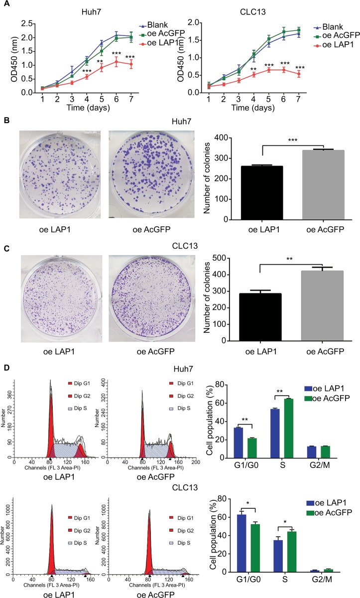
Materials and methods: Differences in LAP1 expression in liver cancer tissues versus matched para-tumoral liver tissues and LCSCs versus non-CSCs were analyzed by Western blotting, real-time polymerase chain reaction, immunohistochemistry, and flow cytometry. The effect of LAP1 on liver cancer cells was evaluated by the expression of CSC markers, oncosphere formation, proliferation, migration, and invasion in vitro. Cell cycle distribution and the number of apoptotic cells were analyzed to assess cell cycle and cell apoptosis. Furthermore, a mouse subcutaneous tumor implant model was established to explore the role of LAP1 in the development of HCC in vivo. Finally, the expression of CSC markers in paraffin-embedded sections was evaluated by immunofluorescence.

Results: LAP1 was weakly expressed in HCC tumors and cell lines and even weaker in LCSCs. LAP1 inhibited the expression of stem cell-associated genes and reduced the abilities of oncosphere formation, proliferation, migration, and invasion in vitro. Cell cycle assay revealed that LAP1 induced G1/G0 arrest. Furthermore, LAP1 decreased subcutaneous tumor-formation ability and the expression of CSC markers and Ki67 in vivo.

Conclusion: LAP1 suppressed the stem cell features of HCC, indicating that it possessed an antitumor effect in liver cancer, both in vitro and in vivo; therefore, LAP1 may prove to be a potential target in liver CSC-targeted therapy.

Keywords: HCC; LAP1; hepatocellular carcinoma; liver cancer stem cell; liver-enriched activator protein 1.


Conflict of interest statement

Disclosure The authors report no conflicts of interest in this work.

Figures

Figure 1
Figure 1
LAP1 is weakly expressed in HCC. Notes: (A) CEBPβ was weakly expressed in patients with HCC. CEBPβ expression in HCC tumor and normal liver tissues provided by Park’s cohort (GSE62232) was analyzed. (B) LAP1 expression levels were verified in samples of patients with HCC by qRT-PCR. GAPDH served as a loading control. (C) HCC samples were assayed by immunohistochemical and H&E staining. Scale bar: 100 µm. (D) The endogenous levels of liver-enriched activating protein and liver-enriched inhibitory protein were determined by Western blot using rabbit IgG anti-CEBPβ (sc-150) and horseradish peroxidase-labeled anti-rabbit IgGs. The α-tubulin protein level was used as a loading control (left panel). For each cell, three independent protein samples were analyzed, and each band was quantified and LAP1 protein expression was normalized with the expression level of the α-tubulin gene. Mean ± SD are represented (right panel). Student’s t-test was used for statistical analysis, *P<0.05 and **P<0.01; data are shown as mean ± SD. Data are representative of at least three independent experiments. Abbreviations: CEBPβ, CCAAT/enhancer binding protein beta; HCC, hepatocellular carcinoma; IgG, immunoglobulin G; LAP1, liver-enriched activator protein 1; qRT-PCR, quantitative real time polymerase chain reaction.
Figure 2
Figure 2
LAP1 is weakly expressed in liver CSC. Notes: (A) LAP1 was weakly expressed in CD13+ cells sorted from Huh7 and CLC13 cells. LAP1 messenger RNA (mRNA) was measured by qRT-PCR. (B) LAP1 was even more weakly expressed in oncospheres than in nonsphere tumor cells; through serial passage of Huh7 and CLC13 sphere cells, similar observations were obtained in the first, second, third, and fourth generations of oncosphere assay. Nonsphere: Huh7 or CLC13 cells that failed to form spheres. Scale bar: 500 µm. Student’s t-test was used for statistical analysis, *P<0.05, **P<0.01, ***P<0.001; data are shown as mean ± SD. Data are representative of at least three independent experiments. Abbreviations: CSC, cancer stem cells; LAP1, liver-enriched activator protein 1; qRT-PCR, quantitative real time polymerase chain reaction.
Figure 3
Figure 3
LAP1 inhibits the stemness of liver CSCs. Notes: (A) LAP1-overexpressing Huh7 cells and control Huh7 cells were established. LAP1-overexpressing CLC13 cells and control CLC13 cells were established (left panel). For each cell, three independent RNA samples were analyzed and CEBPβ mRNA expression was normalized with the expression level of the GAPDH gene. Mean ± SD are represented (right panel). (B) CD13+ (CSC) subpopulations were detected in LAP1-overexpressing Huh7 and LAP1-overexpressing CLC13 cells and their control cells by fluorescence-activated cell sorting (FACS) analysis. Results are shown as mean ± SD. (C) Expression of CD13, CD133, and EpCAM (stemness-associated transcription factors) in LAP1-overexpressing Huh7 and LAP1-overexpressing CLC13 cells, and their control cells were compared by real-time PCR. Significant downregulation in CD13, CD133, and EpCAM mRNA levels was detected in both cell lines after overexpressing LAP1. Results are shown as mean ± SD. (D) LAP1 overexpression causes a diminished oncosphere-forming capacity in Huh7 and CLC13 cells. The right panel represents statistical results as mean ± SD. Scale bar, 200 µm. *P<0.05, **P<0.01, ***P<0.001. Abbreviations: CEBPβ, CCAAT/enhancer binding protein beta; CSC, cancer stem cell; EpCAM, epithelial cell adhesion molecule; LAP1, liver-enriched activator protein 1; oe, overexpression; PCR, polymerase chain reaction.
Figure 4
Figure 4
LAP1 suppress the proliferation of HCC cell line in vitro. Notes: (A) The suppression rate was up to more than 50% on the sixth day after LAP1 infection, compared with AcGFP and noninfected control groups (one-way analysis of variance). *P<0.05, **P<0.01, and ***P<0.001. (B and C) Both HCC cell lines infected with LAP1 formed fewer and smaller colonies than those infected with AcGFP. Each value represents the mean ± SD for triplicate samples (Student’s t-test). *P<0.05, **P<0.01, and ***P<0.001. (D) The cell cycle transition of Huh7 and CLC13 cells was examined, and the increase of the percentage of cells in G1/G0 phase was caused in both cell lines by LAP1 overexpression. All experiments were repeated at least three times, and representative data are shown. Each value represents the mean ± SD for triplicate samples (Student’s t-test). *P<0.05, **P<0.01, and ***P<0.001. Abbreviations: HCC, hepatocellular carcinoma; LAP1, liver-enriched activator protein 1; oe, overexpression.
Figure 5
Figure 5
LAP1 inhibits the migration and invasion of HCC. Notes: (A and B) A wound-healing assay showed that cell motility was suppressed by LAP1 overexpression in Huh7 and CLC13 cells. Microscopic images were acquired at 0, 24, and 48 h after scratching the surface of a confluent layer of cells. (C and D) The in vitro migration ability of Huh7 and CLC13 cells infected with LAP1 was assessed using Transwell assays. Cells infected with AcGFP were used as a control. Scale bar, 200 µm. (E) Matrigel invasion assay was conducted to compare the cell invasion ability between LAP1-overexpressing and control Huh7 cells after 48- and 72-h infection. Invaded cells were fixed and stained with crystal violet, Scale bar, 200 µm. The number of invaded cells was calculated and is shown in the bar chart. All data are mean ± SD of three independent experiments (*P<0.05, **P<0.01, ***P<0.001, Student’s t-test). Abbreviations: LAP1, liver-enriched activator protein; oe, overexpression.
Figure 6
Figure 6
LAP1 suppresses the tumor proliferation and stemness of HCC in vivo. Notes: (A) LAP1-overexpressing and AcGFP-overexpressing Huh7 cells (5×10⁵) and LAP1-overexpressing and AcGFP-overexpressing CLC13 cells (1×10⁶) were subcutaneously injected into BALB/c nude mice for observation of tumor growth. Tumor sizes were observed every 3 days and photographed weekly, and representative pictures are shown. After a 4-week tumor observation, the animals were sacrificed and tumors were removed and weighed. Growth curves of tumors over 4 weeks are summarized and analyzed. Results are shown as mean ± SD, n=6 for each group. (B) The xenograft from the nude mice originated from CLC13 cells were paraffin embedded and IF stained. The level of CEBPβ was monitored using a rabbit CEBPβ antibody, and the overexpression of LAP1 was confirmed. Scale bars: 100 µm. (CE) The level of CD13 or CD133 or Ki67 was compared and analyzed with corresponding antibodies and then counterstained with Hoechst 33342 for confocal microscopy. The images are representative of the results obtained. Scale bars, 100 µm. Abbreviations: CEBPβ, CCAAT/enhancer binding protein beta; HCC, hepatocellular carcinoma; IF, immunofluorescence; LAP1, liver-enriched activator protein 1; oe, overexpression.

Similar articles

Cited by

References

    1. Torre LA, Bray F, Siegel RL, Ferlay J, Lortet-Tieulent J, Jemal A. Global cancer statistics, 2012. CA Cancer J Clin. 2015;65(2):87–108. - PubMed
    1. Wang R, Sun Q, Wang P, et al. Notch and Wnt/β-catenin signaling pathway play important roles in activating liver cancer stem cells. Oncotarget. 2016;7(5):5754–5768. - PMC - PubMed
    1. Siegel RL, Miller KD, Jemal A. Cancer statistics, 2016. CA Cancer J Clin. 2016;66(1):7–30. - PubMed
    1. Maluccio M, Covey A. Recent progress in understanding, diagnosing, and treating hepatocellular carcinoma. CA Cancer J Clin. 2012;62(6):394–399. - PubMed
    1. Hao J, Zhang Y, Deng M, et al. MicroRNA control of epithelial-mesenchymal transition in cancer stem cells. Int J Cancer. 2014;135(5):1019–1027. - PubMed