. 2018 Apr;27(2):120-128.
doi: 10.5607/en.2018.27.2.120. Epub 2018 Apr 24.

Expression of µ-Opioid Receptor in CA1 Hippocampal Astrocytes

Affiliations

Expression of µ-Opioid Receptor in CA1 Hippocampal Astrocytes

Min-Ho Nam et al. Exp Neurobiol. 2018 Apr.

Abstract

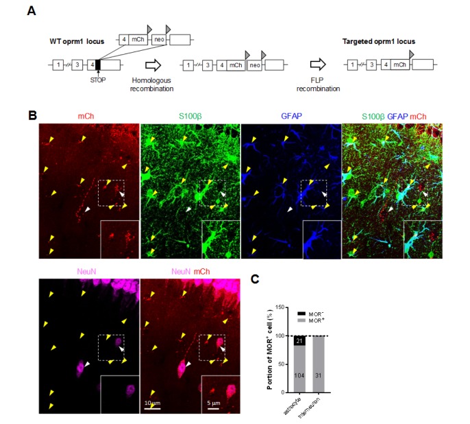
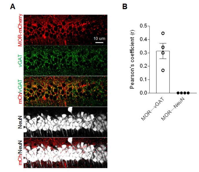
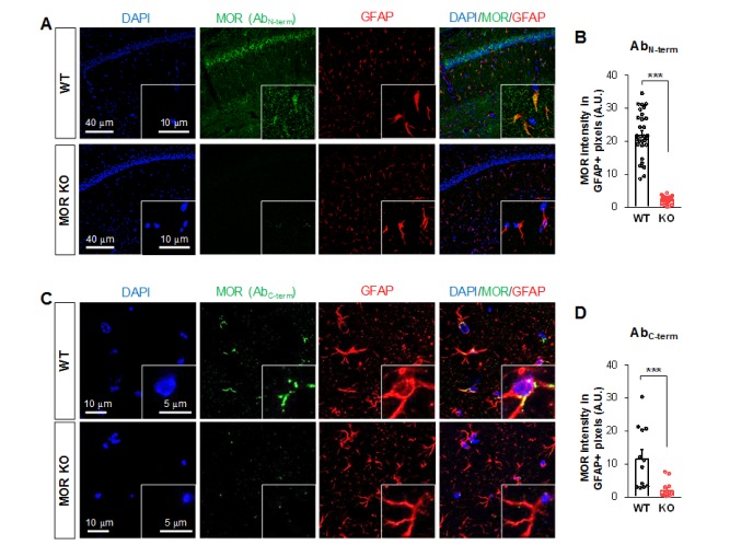
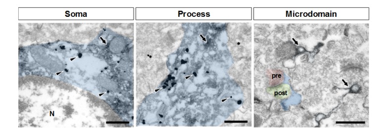
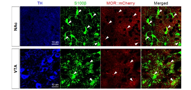
µ-opioid receptor (MOR) is a class of opioid receptors with a high affinity for enkephalins and beta-endorphin. In hippocampus, activation of MOR is known to enhance the neuronal excitability of pyramidal neurons, which has been mainly attributed to a disinhibition of pyramidal neurons via activating Gαi subunit to suppress the presynaptic release of GABA in hippocampal interneurons. In contrast, the potential role of MOR in hippocampal astrocytes, the most abundant cell type in the brain, has remained unexplored. Here, we determine the cellular and subcellular distribution of MOR in different cell types of the hippocampus by utilizing MOR-mCherry mice and two different antibodies against MOR. Consistent with previous findings, we demonstrate that MOR expression in the CA1 pyramidal layer is co-localized with axon terminals from GABAergic inhibitory neurons but not with soma of pyramidal neurons. More importantly, we demonstrate that MOR is highly expressed in CA1 hippocampal astrocytes. The ultrastructural analysis further demonstrates that the astrocytic MOR is localized in soma and processes, but not in microdomains near synapses. Lastly, we demonstrate that astrocytes in ventral tegmental area and nucleus accumbens also express MOR. Our results provide the unprecedented evidence for the presence of MOR in astrocytes, implicating potential roles of astrocytic MOR in addictive behaviors.

Keywords: Astrocyte; Electron microscopy; Hippocampus; µ-opioid receptor.


Figures

Fig. 1
Fig. 1. Expression of MOR in CA1 hippocampal interneurons and astrocytes in MOR-mCherry mice. (A) Genetic background of MOR-mCherry knock-in mouse. Triangle, FRT; mCh, mcherry cDNA; neo, flanked neomycin cassette. (B) Expression of MOR-mCherry signal in GFAP+/S100β+ astrocytes (yellow arrowheads) and NeuN+ interneurons (white arrowheads) in hippocampal CA1 stratum radiatum of MOR-mCherry mouse. (C) Quantification of the portion of MOR+ astrocytes and MOR+ interneurons. Numbers on the bar graph indicate the cell numbers.
Fig. 2
Fig. 2. Colocalization between MOR and vGAT in CA1 pyramidal layer. (A) Representative confocal images. (B) Pearson's coefficient of colocalizations of both MOR-mCherry - vGAT and MOR-mCherry - NeuN measured by Colocalization Threshold tool from ImageJ.
Fig. 3
Fig. 3. Expression of MOR in hippocampal astrocytes immunostained by two different antibodies. (A, C) Representative confocal images of the expression of MOR (green; antibodies targeting amino acids 1~80 mapping near the N-terminus (A) or 1359~1403 of the C-terminus of MOR (C)) in GFAP-positive astrocytes (red) of hippocampal CA1 area. (B, D) Bar graph showing the comparison of the average pixel intensity of MOR immunoreactivity signal in GFAP-positive cells of WT and MOR KO mice. Numbers in the bar graph indicate examined cells from 3 mice for each group. Unpaired two-tailed t-test (**p<0.01, ***p<0.001).
Fig. 4
Fig. 4. Validation of antibody using MOR shRNA. (A) Confocal images of MOR (green) after infection of lentivirus carrying MOR-shRNA-katushka (upper) or scrambled-shRNA-katushka (lower), respectively. (B) In vitro knockdown efficiency test of MOR shRNA in cultured hippocampal astrocytes by western blot. Upper band indicates MOR immunoactivity and lower band indicates actin immunoactivity. (C) In vivo knockdown efficiency test of MOR shRNA in CA1 hippocampus by western blot.
Fig. 5
Fig. 5. Subcellular distribution (soma, process, and microdomain) of MOR in astrocyte (indicated in blue). MOR is stained with immunogold with silver enhancement (dark specks, arrowheads), and GFP, representing astrocyte, is stained with immunoperoxidase (dark amorphous deposits, arrows). The soma, process, and microdomain of the astrocyte were colored blue. Presynaptic axon terminal (pre) and postsynaptic dendrite (post) were colored red and green, respectively. N is nucleus. Scale bar indicates 500 nm.
Fig. 6
Fig. 6. MOR expression in S100β-positive astrocytes in mesolimbic area. S100β+ astrocytes in NAc and VTA which were marked by tyrosine hydroxylase (TH) express MOR-mCherry signals. Arrowheads indicate MOR-mcherry+/S100β+ astrocytes.

References

    1. Saidak Z, Blake-Palmer K, Hay DL, Northup JK, Glass M. Differential activation of G-proteins by mu-opioid receptor agonists. Br J Pharmacol. 2006;147:671–680. - PMC - PubMed
    1. Arvidsson U, Riedl M, Chakrabarti S, Lee JH, Nakano AH, Dado RJ, Loh HH, Law PY, Wessendorf MW, Elde R. Distribution and targeting of a mu-opioid receptor (MOR1) in brain and spinal cord. J Neurosci. 1995;15:3328–3341. - PMC - PubMed
    1. Garzón M, Pickel VM. Plasmalemmal mu-opioid receptor distribution mainly in nondopaminergic neurons in the rat ventral tegmental area. Synapse. 2001;41:311–328. - PubMed
    1. Svoboda KR, Adams CE, Lupica CR. Opioid receptor subtype expression defines morphologically distinct classes of hippocampal interneurons. J Neurosci. 1999;19:85–95. - PMC - PubMed
    1. Drake CT, Milner TA. Mu opioid receptors are in somatodendritic and axonal compartments of GABAergic neurons in rat hippocampal formation. Brain Res. 1999;849:203–215. - PubMed