. 2018 Apr 20:9:686.
doi: 10.3389/fimmu.2018.00686. eCollection 2018.

Adaptive NKG2C+CD57+ Natural Killer Cell and Tim-3 Expression During Viral Infections

Affiliations

Adaptive NKG2C+CD57+ Natural Killer Cell and Tim-3 Expression During Viral Infections

Hassen Kared et al. Front Immunol. .

Abstract

Repetitive stimulation by persistent pathogens such as human cytomegalovirus (HCMV) or human immunodeficiency virus (HIV) induces the differentiation of natural killer (NK) cells. This maturation pathway is characterized by the acquisition of phenotypic markers, CD2, CD57, and NKG2C, and effector functions-a process regulated by Tim-3 and orchestrated by a complex network of transcriptional factors, involving T-bet, Eomes, Zeb2, promyelocytic leukemia zinc finger protein, and Foxo3. Here, we show that persistent immune activation during chronic viral co-infections (HCMV, hepatitis C virus, and HIV) interferes with the functional phenotype of NK cells by modulating the Tim-3 pathway; a decrease in Tim-3 expression combined with the acquisition of inhibitory receptors skewed NK cells toward an exhausted and cytotoxic phenotype in an inflammatory environment during chronic HIV infection. A better understanding of the mechanisms underlying NK cell differentiation could aid the identification of new immunological targets for checkpoint blockade therapies in a manner that is relevant to chronic infection and cancer.

Keywords: aging; cancer; checkpoint blockade; chronic infection; exhaustion; maturation; natural killer cells; senescence.


Figures
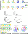
Figure 1
Figure 1
Identification of adaptive natural killer (NK) cells during human cytomegalovirus (HCMV) infection. (A) Identification of HCMV-induced human NK cells by mass cytometry. Frozen PBMCs from HCMV-seronegative and -seropositive donors were surface and intracellular stained for mass cytometry analysis. Samples were barcoded and acquired simultaneously (n = 6). t-Distributed stochastic neighbor embedding (tSNE) analysis of 23 parametric data was performed on live CD45+CD14CD19CD3CD56+ NK cells from three HCMV-seropositive and three HCMV-seronegative donors. Event density in the tSNE field for all donors compiled together in which a same number of events per donor was included. Normalized protein expression levels for CD56, CD16, CD94, NKG2C, and CD57 in tSNE field were represented in red for high expression, whereas blue represents low expression (cold-to-hot heat map). (B) The antiviral response against HCMV drives acquisition of CD85j and NKG2C in CD57+ NK cells. The frequency and phenotype of NK cells subsets were analyzed by flow cytometry on freshly isolated PBMC (n = 28). The frequency of NKG2C+Siglec-7 and CD85j+CD57+CD16+CD56dim NK cells were positively correlated with the level of IgG antibodies specific to HCMV (p = 0.0009; r = 0.6832 and p = 0.0199; r = 0.5158, respectively). (C) Identification of human NKG2C+ adaptive NK cells by flow cytometry. PBMCs from HCMV-seronegative and -seropositive donors were surface and intracellular stained for flow cytometry analysis. Samples were acquired individually (n = 24). tSNE analysis of 18 parametric data was performed on live lymphocytes CD45+CD14CD19CD3CD56+ NK cells from 12 HCMV-seropositive and 12 HCMV-seronegative donors. Event density in the tSNE field for all donors compiled together in which 5,000 events per donor were included. Normalized protein expression levels for single parameters in tSNE field were represented with a cold-to-hot heat map. (D) Identification of human NKG2C+ adaptive NK cells clusters during HCMV infection. HCMV-seropositive and -seronegative donors were compiled separately and analyzed for the repartition of clusters. (E) Phenotype of human NKG2C+ adaptive NK cells in presence or absence of HCMV infection. Gated NKG2C+CD56dim and NKG2CCD56dim NK cells from 12 HCMV-seropositive and 12 HCMV-seronegative donors were analyzed by flow cytometry to detect specific signature of adaptive NK cells through the surface and intracellular expression of markers previously described as associated with HCMV infection. Each donor was identified by a unique symbol for the different molecules studied. The median fluorescent intensity or frequency of markers expression was represented for molecules with continuous or bi-modal expression, respectively. The median values were compared using a Wilcoxon matched-pairs signed rank test (*p < 0.05, **p < 0.01, ***p < 0.001, and ****p < 0.0001). (F) Characterization of human NK cell’s maturation according to Wanderlust trajectory. The Wanderlust trajectory is fixed to an arbitrary scale where the most immature NK cells (CD56bright) are at 0 and the most mature at 1. The traces demonstrated the relative expression patterns of Siglec-7, CD62L, CD57, NKG2C, CD85j, Ceacam-1, and Tim-3 across differentiation in HCMV-seropositive and HCMV-seronegative donors. (G,H) The expression of proteins and Wanderlust scale were normalized for the same markers than in panel (F) or for a new set of antigens such as NKG2A, NKG2D, CD38, CD161, and transcription factors promyelocytic leukemia zinc finger protein, Helios, Eomes, and T-bet. NKG2C and CD2 staining were included in the analysis but data are not shown. The variance of proteins expression was represented to illustrate heterogeneity between HCMV-seropositive donors.
Figure 1
Figure 1
Identification of adaptive natural killer (NK) cells during human cytomegalovirus (HCMV) infection. (A) Identification of HCMV-induced human NK cells by mass cytometry. Frozen PBMCs from HCMV-seronegative and -seropositive donors were surface and intracellular stained for mass cytometry analysis. Samples were barcoded and acquired simultaneously (n = 6). t-Distributed stochastic neighbor embedding (tSNE) analysis of 23 parametric data was performed on live CD45+CD14CD19CD3CD56+ NK cells from three HCMV-seropositive and three HCMV-seronegative donors. Event density in the tSNE field for all donors compiled together in which a same number of events per donor was included. Normalized protein expression levels for CD56, CD16, CD94, NKG2C, and CD57 in tSNE field were represented in red for high expression, whereas blue represents low expression (cold-to-hot heat map). (B) The antiviral response against HCMV drives acquisition of CD85j and NKG2C in CD57+ NK cells. The frequency and phenotype of NK cells subsets were analyzed by flow cytometry on freshly isolated PBMC (n = 28). The frequency of NKG2C+Siglec-7 and CD85j+CD57+CD16+CD56dim NK cells were positively correlated with the level of IgG antibodies specific to HCMV (p = 0.0009; r = 0.6832 and p = 0.0199; r = 0.5158, respectively). (C) Identification of human NKG2C+ adaptive NK cells by flow cytometry. PBMCs from HCMV-seronegative and -seropositive donors were surface and intracellular stained for flow cytometry analysis. Samples were acquired individually (n = 24). tSNE analysis of 18 parametric data was performed on live lymphocytes CD45+CD14CD19CD3CD56+ NK cells from 12 HCMV-seropositive and 12 HCMV-seronegative donors. Event density in the tSNE field for all donors compiled together in which 5,000 events per donor were included. Normalized protein expression levels for single parameters in tSNE field were represented with a cold-to-hot heat map. (D) Identification of human NKG2C+ adaptive NK cells clusters during HCMV infection. HCMV-seropositive and -seronegative donors were compiled separately and analyzed for the repartition of clusters. (E) Phenotype of human NKG2C+ adaptive NK cells in presence or absence of HCMV infection. Gated NKG2C+CD56dim and NKG2CCD56dim NK cells from 12 HCMV-seropositive and 12 HCMV-seronegative donors were analyzed by flow cytometry to detect specific signature of adaptive NK cells through the surface and intracellular expression of markers previously described as associated with HCMV infection. Each donor was identified by a unique symbol for the different molecules studied. The median fluorescent intensity or frequency of markers expression was represented for molecules with continuous or bi-modal expression, respectively. The median values were compared using a Wilcoxon matched-pairs signed rank test (*p < 0.05, **p < 0.01, ***p < 0.001, and ****p < 0.0001). (F) Characterization of human NK cell’s maturation according to Wanderlust trajectory. The Wanderlust trajectory is fixed to an arbitrary scale where the most immature NK cells (CD56bright) are at 0 and the most mature at 1. The traces demonstrated the relative expression patterns of Siglec-7, CD62L, CD57, NKG2C, CD85j, Ceacam-1, and Tim-3 across differentiation in HCMV-seropositive and HCMV-seronegative donors. (G,H) The expression of proteins and Wanderlust scale were normalized for the same markers than in panel (F) or for a new set of antigens such as NKG2A, NKG2D, CD38, CD161, and transcription factors promyelocytic leukemia zinc finger protein, Helios, Eomes, and T-bet. NKG2C and CD2 staining were included in the analysis but data are not shown. The variance of proteins expression was represented to illustrate heterogeneity between HCMV-seropositive donors.
Figure 2
Figure 2
Regulation of adaptive natural killer (NK) cells by Tim-3 pathway during human cytomegalovirus (HCMV) infection. (A) Increased IFN-γ secretion is a hallmark of adaptive NK cells. Frozen PBMCs from 10 HCMV-seropositive donors were stimulated overnight with cytokines (IL-12/IL-15/IL-18) or by the contact of K562 cells. Surface and intracellular flow cytometry staining was performed to characterize the phenotype (Tim-3, Eomes) and functions (IFN-γ) of conventional (identified as CD45+CD14CD19CD3CD56+NKG2C) and adaptive (CD45+CD14CD19CD3CD56+NKG2C+) live NK cells. (B) Increased IFN-γ secretion by adaptive NK cells is associated with enhancement of Tim-3 and Eomes expression. The median fluorescence intensity of CD2, Tim-3, and Eomes was measured in NKG2C+ NK cells before/after stimulation by inflammatory cytokines or interaction with cancer cell lines lacking MHC I expression (except Tim-3). The release of IFN-γ, TNF-α, and cytotoxicity (CD107a+GZB+) by gated NKG2C and NKG2C+ NK cells was assessed after stimulation with IL-12/IL-15/IL-18 (10/20/100 ng/ml, respectively) or interaction with K562 cell lines by flow cytometry. (C) Identification of polyfunctional NK cells using CD57 and NKG2C expression. CD56dim NK cells from HCMV-seropositive donors were sorted according to CD57 and NKG2C expression and stimulated by CD16 crosslinking. Supernatants were collected and analyzed by Luminex. The heat map represented the median concentration of each molecule. (D) Regulation of CD57+NKG2C+ NK cells polyfunctionality by Tim-3 expression. Sorted NK cell subsets were preincubated with anti-Tim-3 (10 µg/ml) or IgG control before overnight stimulation by CD16 ligation. Supernatants of stimulated NK cells subsets were analyzed by Luminex, and the median concentrations of each analyte were represented by heat map. (E) Upregulation of Zeb2 expression in CD57+NKG2C+ NK cells and differential expression of Foxo3 and TBX21 during NK cell maturation. Sorted NK and T cell subsets from HCMV-seropositive donors were immediately lysed. Senescent T cells (CD57) and immature NK cells (CD56bright) were used as internal control. Transcription factor expression was analyzed by RT-PCR and normalized according to the expression of actin B used as a housekeeping gene. (F) T-bet and Foxo3 expression but not Zeb2 CD57+NKG2C+ NK cells are regulated by Tim-3 expression. Sorted NK cell subsets from HCMV-seropositive donors were preincubated with anti-Tim-3 or IgG control before overnight stimulation by CD16 ligation. Gene expressions of NK cell subsets were analyzed directly ex vivo and after in vitro stimulation with CD16 stimulation in presence of blocking Tim-3 antibody or IgG control. (G) Ceacam-1 silencing interferes with NK cell functions. Frozen PBMC from six HCMV-seropositive donors were pre-activated overnight with IL-2 and IL-15 before transfection with scrambled or specific Ceacam-1 siRNA. After resting, transfected cells were stimulated with CD16 crosslinking. CD57+ NK cells are depicted in black (Scr) or red (Ceacam-1) lines, and CD57 NK cells are depicted in blue (Scr) or green (Ceacam-1) lines. The intensities of Ki-67 and T-bet were measured by intracellular staining on gated CD3CD56dimCD16+CD57 and CD3CD56dimCD16+CD57+ NK cells. (H) Ceacam-1 silencing induces NK cell proliferation and T-bet expression. Ki-67 and T-bet expressions have been measured by flow cytometry after transfection with control scrambled (Scr) or specific Ceacam-1 siRNA. (I) Tim-3 expression regulates susceptibility of adaptive NK cells to apoptosis induced by Galectin-9. PBMCs of 10 HCMV-seropositive donors were stimulated by CD16 ligation in the presence of recombinant Galectin-9 (1 µM). Apoptosis was measured by the staining of annexin-V combined with 7-AAD staining on gated CD3CD56dimCD16+ NK cells segregated according to expression of CD57, Tim-3 (J) or NKG2C (K). Unstimulated cells and PBMCs incubated with Staurosporine were used, respectively, as a negative and positive control for apoptosis and necrosis. The mean values were compared using a paired t-test (*p < 0.05 and **p < 0.01).
Figure 2
Figure 2
Regulation of adaptive natural killer (NK) cells by Tim-3 pathway during human cytomegalovirus (HCMV) infection. (A) Increased IFN-γ secretion is a hallmark of adaptive NK cells. Frozen PBMCs from 10 HCMV-seropositive donors were stimulated overnight with cytokines (IL-12/IL-15/IL-18) or by the contact of K562 cells. Surface and intracellular flow cytometry staining was performed to characterize the phenotype (Tim-3, Eomes) and functions (IFN-γ) of conventional (identified as CD45+CD14CD19CD3CD56+NKG2C) and adaptive (CD45+CD14CD19CD3CD56+NKG2C+) live NK cells. (B) Increased IFN-γ secretion by adaptive NK cells is associated with enhancement of Tim-3 and Eomes expression. The median fluorescence intensity of CD2, Tim-3, and Eomes was measured in NKG2C+ NK cells before/after stimulation by inflammatory cytokines or interaction with cancer cell lines lacking MHC I expression (except Tim-3). The release of IFN-γ, TNF-α, and cytotoxicity (CD107a+GZB+) by gated NKG2C and NKG2C+ NK cells was assessed after stimulation with IL-12/IL-15/IL-18 (10/20/100 ng/ml, respectively) or interaction with K562 cell lines by flow cytometry. (C) Identification of polyfunctional NK cells using CD57 and NKG2C expression. CD56dim NK cells from HCMV-seropositive donors were sorted according to CD57 and NKG2C expression and stimulated by CD16 crosslinking. Supernatants were collected and analyzed by Luminex. The heat map represented the median concentration of each molecule. (D) Regulation of CD57+NKG2C+ NK cells polyfunctionality by Tim-3 expression. Sorted NK cell subsets were preincubated with anti-Tim-3 (10 µg/ml) or IgG control before overnight stimulation by CD16 ligation. Supernatants of stimulated NK cells subsets were analyzed by Luminex, and the median concentrations of each analyte were represented by heat map. (E) Upregulation of Zeb2 expression in CD57+NKG2C+ NK cells and differential expression of Foxo3 and TBX21 during NK cell maturation. Sorted NK and T cell subsets from HCMV-seropositive donors were immediately lysed. Senescent T cells (CD57) and immature NK cells (CD56bright) were used as internal control. Transcription factor expression was analyzed by RT-PCR and normalized according to the expression of actin B used as a housekeeping gene. (F) T-bet and Foxo3 expression but not Zeb2 CD57+NKG2C+ NK cells are regulated by Tim-3 expression. Sorted NK cell subsets from HCMV-seropositive donors were preincubated with anti-Tim-3 or IgG control before overnight stimulation by CD16 ligation. Gene expressions of NK cell subsets were analyzed directly ex vivo and after in vitro stimulation with CD16 stimulation in presence of blocking Tim-3 antibody or IgG control. (G) Ceacam-1 silencing interferes with NK cell functions. Frozen PBMC from six HCMV-seropositive donors were pre-activated overnight with IL-2 and IL-15 before transfection with scrambled or specific Ceacam-1 siRNA. After resting, transfected cells were stimulated with CD16 crosslinking. CD57+ NK cells are depicted in black (Scr) or red (Ceacam-1) lines, and CD57 NK cells are depicted in blue (Scr) or green (Ceacam-1) lines. The intensities of Ki-67 and T-bet were measured by intracellular staining on gated CD3CD56dimCD16+CD57 and CD3CD56dimCD16+CD57+ NK cells. (H) Ceacam-1 silencing induces NK cell proliferation and T-bet expression. Ki-67 and T-bet expressions have been measured by flow cytometry after transfection with control scrambled (Scr) or specific Ceacam-1 siRNA. (I) Tim-3 expression regulates susceptibility of adaptive NK cells to apoptosis induced by Galectin-9. PBMCs of 10 HCMV-seropositive donors were stimulated by CD16 ligation in the presence of recombinant Galectin-9 (1 µM). Apoptosis was measured by the staining of annexin-V combined with 7-AAD staining on gated CD3CD56dimCD16+ NK cells segregated according to expression of CD57, Tim-3 (J) or NKG2C (K). Unstimulated cells and PBMCs incubated with Staurosporine were used, respectively, as a negative and positive control for apoptosis and necrosis. The mean values were compared using a paired t-test (*p < 0.05 and **p < 0.01).
Figure 3
Figure 3
Tim-3 expression and adaptive natural killer (NK) cells during human immunodeficiency virus (HIV) infection. (A) Induction of CD57+NKG2C+ NK cells during HIV therapy. Untreated HIV patients were followed longitudinally before and after the initiation of antiretroviral treatment (48 weeks). Representative zebra plots of adaptive markers and inhibitory receptors expression on total NK cells (CD3negCD56+) during highly active antiretroviral therapy (HAART) follow-up. (B) Inhibitory receptors expression of mature NK cells during HIV infection. Fifteen HIV infected patients were monitored before and after HIV treatment. Tim-3, T-bet, and Eomes modulation in NK cells were not statistically significant modulated by HAART. The median values were compared using a Wilcoxon matched-pairs signed rank test (*p < 0.05, **p < 0.01, ***p < 0.001, and ****p < 0.0001). (C) Downmodulation of Tim-3 but preservation of NKG2C on CD57+ NK cells during HIV infection. The frequencies of Tim-3 and NKG2C in CD57 and CD57+NK cells (gated as viable CD56+CD3neg cells) were studied in our cross-sectional HIV cohort (Malaysia) and compared with healthy donors from Singapore (first column) or Malaysia (second column). (D) Increased expression of inhibitory receptors and transcription factor (TF) expression in CD57+ NK cells during HIV infection. The frequencies of inhibitory receptors and TF in CD57+ NK cells (gated as viable CD56+CD3neg cells) were normalized to obtain a mean row Z-score for each marker. A cold-to-hot heat map represented the relative expression of molecules in each patient group (age did not impact on the expression of most of these molecules and was not included here to simplify the representation). (E) Tim-3 expression, immune activation, and inhibitory receptors. Soluble CD14 correlated negatively with Tim-3 expression on mature CD57+ NK cells from HIV-infected and HIV-non-infected individuals (n = 119). The loss of Tim-3 expression is also coupled with acquisition of Ceacam-1 on CD57+ NK cells from HIV-infected and HIV-non-infected individuals (n = 128). The acquisition of Tim-3 was positively associated with T-bet (n = 145) and inhibitory receptors including CD160 (n = 145) and T-cell immuno-receptor with Ig and ITIM domain (TIGIT) (n = 145) in all donors. We used the non-parametric Spearman rank-order test to compare correlation between Tim-3 and sCD14 or proteins expression on CD57+ NK cells. We reported r-values and p-values. Analysis with *p < 0.05, **p < 0.01, and ***p < 0.001 were considered significantly different between the groups. (F) NK cells functions and inhibitory receptors expression during HIV infection. Representation of a combined phenotype and functional assay in total NK cells from non-infected and HIV infected patients. (G) Differential role of Tim-3 acquisition during NK cells activation in healthy donors or (H) during chronic HIV infection. Tim-3 or TNF-α secretion are positively correlated with IFN-γ intensity in NK cells from healthy donors. The inhibitory receptors TIGIT and Tim-3 are negatively correlated with IFN-γ secretion by NK cells in HAART-treated HIV patients.
Figure 3
Figure 3
Tim-3 expression and adaptive natural killer (NK) cells during human immunodeficiency virus (HIV) infection. (A) Induction of CD57+NKG2C+ NK cells during HIV therapy. Untreated HIV patients were followed longitudinally before and after the initiation of antiretroviral treatment (48 weeks). Representative zebra plots of adaptive markers and inhibitory receptors expression on total NK cells (CD3negCD56+) during highly active antiretroviral therapy (HAART) follow-up. (B) Inhibitory receptors expression of mature NK cells during HIV infection. Fifteen HIV infected patients were monitored before and after HIV treatment. Tim-3, T-bet, and Eomes modulation in NK cells were not statistically significant modulated by HAART. The median values were compared using a Wilcoxon matched-pairs signed rank test (*p < 0.05, **p < 0.01, ***p < 0.001, and ****p < 0.0001). (C) Downmodulation of Tim-3 but preservation of NKG2C on CD57+ NK cells during HIV infection. The frequencies of Tim-3 and NKG2C in CD57 and CD57+NK cells (gated as viable CD56+CD3neg cells) were studied in our cross-sectional HIV cohort (Malaysia) and compared with healthy donors from Singapore (first column) or Malaysia (second column). (D) Increased expression of inhibitory receptors and transcription factor (TF) expression in CD57+ NK cells during HIV infection. The frequencies of inhibitory receptors and TF in CD57+ NK cells (gated as viable CD56+CD3neg cells) were normalized to obtain a mean row Z-score for each marker. A cold-to-hot heat map represented the relative expression of molecules in each patient group (age did not impact on the expression of most of these molecules and was not included here to simplify the representation). (E) Tim-3 expression, immune activation, and inhibitory receptors. Soluble CD14 correlated negatively with Tim-3 expression on mature CD57+ NK cells from HIV-infected and HIV-non-infected individuals (n = 119). The loss of Tim-3 expression is also coupled with acquisition of Ceacam-1 on CD57+ NK cells from HIV-infected and HIV-non-infected individuals (n = 128). The acquisition of Tim-3 was positively associated with T-bet (n = 145) and inhibitory receptors including CD160 (n = 145) and T-cell immuno-receptor with Ig and ITIM domain (TIGIT) (n = 145) in all donors. We used the non-parametric Spearman rank-order test to compare correlation between Tim-3 and sCD14 or proteins expression on CD57+ NK cells. We reported r-values and p-values. Analysis with *p < 0.05, **p < 0.01, and ***p < 0.001 were considered significantly different between the groups. (F) NK cells functions and inhibitory receptors expression during HIV infection. Representation of a combined phenotype and functional assay in total NK cells from non-infected and HIV infected patients. (G) Differential role of Tim-3 acquisition during NK cells activation in healthy donors or (H) during chronic HIV infection. Tim-3 or TNF-α secretion are positively correlated with IFN-γ intensity in NK cells from healthy donors. The inhibitory receptors TIGIT and Tim-3 are negatively correlated with IFN-γ secretion by NK cells in HAART-treated HIV patients.

References

    1. Townsend MJ, Weinmann AS, Matsuda JL, Salomon R, Farnham PJ, Biron CA, et al. T-bet regulates the terminal maturation and homeostasis of NK and Valpha14i NKT cells. Immunity (2004) 20(4):477–94. 10.1016/S1074-7613(04)00076-7 - DOI - PubMed
    1. Lopez-Verges S, Milush JM, Schwartz BS, Pando MJ, Jarjoura J, York VA, et al. Expansion of a unique CD57(+)NKG2Chi natural killer cell subset during acute human cytomegalovirus infection. Proc Natl Acad Sci U S A (2011) 108(36):14725–32. 10.1073/pnas.1110900108 - DOI - PMC - PubMed
    1. Bjorkstrom NK, Riese P, Heuts F, Andersson S, Fauriat C, Ivarsson MA, et al. Expression patterns of NKG2A, KIR, and CD57 define a process of CD56dim NK-cell differentiation uncoupled from NK-cell education. Blood (2010) 116(19):3853–64. 10.1182/blood-2010-04-281675 - DOI - PubMed
    1. Della Chiesa M, Falco M, Bertaina A, Muccio L, Alicata C, Frassoni F, et al. Human cytomegalovirus infection promotes rapid maturation of NK cells expressing activating killer Ig-like receptor in patients transplanted with NKG2C-/- umbilical cord blood. J Immunol (2014) 192(4):1471–9. 10.4049/jimmunol.1302053 - DOI - PubMed
    1. Lopez-Verges S, Milush JM, Pandey S, York VA, Arakawa-Hoyt J, Pircher H, et al. CD57 defines a functionally distinct population of mature NK cells in the human CD56dimCD16+ NK-cell subset. Blood (2010) 116(19):3865–74. 10.1182/blood-2010-04-282301 - DOI - PMC - PubMed

Publication types

MeSH terms

Substances