. 2018 May;15(5):4191-4198.
doi: 10.3892/etm.2018.5973. Epub 2018 Mar 20.

Methotrexate remediates spinal cord injury in vivo and in vitro via suppression of endoplasmic reticulum stress-induced apoptosis

Affiliations

Methotrexate remediates spinal cord injury in vivo and in vitro via suppression of endoplasmic reticulum stress-induced apoptosis

Fengju Rong et al. Exp Ther Med. 2018 May.

Abstract

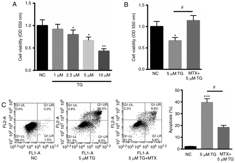
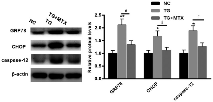
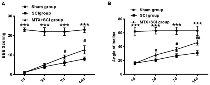
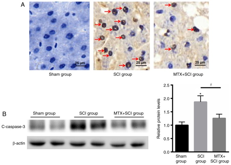
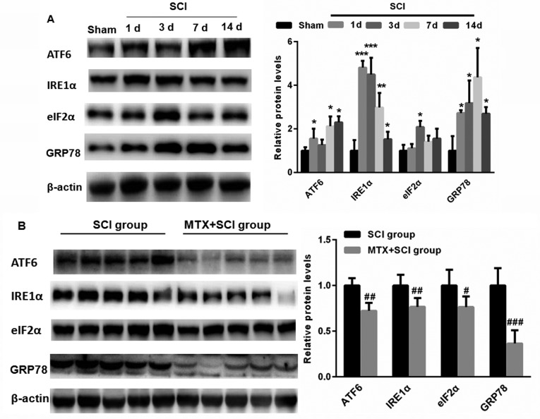
It has been suggested that endoplasmic reticulum stress (ERS) may induce apoptosis following spinal cord injury (SCI). Methotrexate (MTX) has been used as a long-term therapy regimen for rheumatoid arthritis. However, it is not clear whether MTX remediates SCI by inhibiting ERS. In the present study, to establish an in vitro ERS cell model, PC12 cells were pre-incubated with triglycerides (TG). MTT assays revealed that treatment with 1, 2.5, 5 and 10 µM TG decreased PC12 cell viability in a dose-dependent manner. Additionally, MTX treatment significantly reversed the TG-induced decrease in cell viability and increased apoptosis according to the flow cytometry assay (P<0.05). Notably, western blotting indicated that MTX significantly decreased levels of glucose-regulated protein (GRP)78, CCAAT-enhancer-binding protein homologous protein (CHOP) and caspase-12 expression (P<0.05), which were increased following treatment with TG. Furthermore, the in vivo role of MTX in a rat model of SCI was evaluated. The motor behavioral function of rats was improved following treatment with MTX according to Basso, Beattie and Bresnahan scoring (P<0.05). Terminal deoxynucleotidyl-transferase-mediated dUTP nick end staining indicated that there were no apoptotic cells present in sham rats. In the SCI model group, apoptotic cells were observed at day 7; however, the number of apoptotic cells was reduced following an additional 7 days of MTX administration. Furthermore, levels of ERS-associated proteins, including caspase-3, activating transcription factor 6, serine/threonine-protein kinase/endoribonuclease inositol-requiring enzyme 1 α, eukaryotic initiation factor 2 α and GRP78, were significantly increased following SCI; however, administration of MTX for 7 days significantly reversed this effect (P<0.05, P<0.01 and P<0.001). Therefore, MTX may improve SCI by suppressing ERS-induced apoptosis in vitro and in vivo.

Keywords: apoptosis; endoplasmic reticulum stress; methotrexate; spinal cord injury.


Figures

Figure 1.
Figure 1.
MTX inhibited TG-induced PC12 cell apoptosis. (A) MTT assay of PC12 cells treated with 1, 2.5, 5, and 10 µM TG. (B) MTX treatment significantly reversed the TG-induced decrease in cell viability by 46.7%. (C) Flow cytometry analysis of PC12 cells treated with 5 µM TG in the presence or absence of MTX. FL1-A on the X-axis represents the relative fluorescence intensity value; FL2-A on the Y-axis represents the relative fluorescence intensity value. Data are presented as the mean ± standard error of the mean. *P<0.05, **P<0.01 and ***P<0.001 vs. NC; #P<0.05. NC, normal control; MTX, methotrexate; TG, triglycerides; OD, optical density.
Figure 2.
Figure 2.
MTX reversed the effects of TG on the levels of GRP78, CHOP and caspase-12 expression in PC12 cells. Western blotting was performed to assess the levels of GRP78, CHOP and caspase-12 expression in PC12 cells treated with 5 µM TG in the presence or absence of MTX. Data are presented as the mean ± standard error of the mean. *P<0.05 and **P<0.01 vs. NC; #P<0.05. GRP78, glucose-regulated protein 78; CHOP, CCAAT-enhancer-binding protein homologous protein; NC, normal control (dimethyl sulfoxide); MTX, methotrexate; TG, triglycerides; NC, normal control.
Figure 3.
Figure 3.
MTX improved motor behavioral function in rats with SCI. (A) BBB scoring and the (B) inclined plate test were performed on rats in the sham, SCI and MTX+SCI groups (n=8 rats per group). Data are presented as the mean ± standard error of the mean. ***P<0.001 vs. Sham group; #P<0.05 and ##P<0.01 vs. SCI group. BBB, Basso, Beattie and Bresnahan; SCI, spinal cord injury; MTX, methotrexate.
Figure 4.
Figure 4.
MTX reduced apoptosis in rats with SCI. (A) Terminal deoxynucleotidyl-transferase-mediated dUTP nick end staining (magnification, ×40) indicated that the sham group demonstrated no obvious cell apoptosis with blue staining, whereas an increased number of apoptotic cells in SCI group on day 7 and improvement of cell apoptosis after MTX treatment on day 7 were identified (TUNEL-positive nuclei were stained brown and TUNEL-negative nuclei were stained blue; red arrows indicated apoptotic cells). (B) Western blotting of c-caspase-3 protein expression in the sham, SCI and MTX+SCI group (n=8 rats per group). Data are presented as the mean ± standard error of the mean. *P<0.05 vs. Sham; #P<0.05. SCI, spinal cord injury; MTX, methotrexate; c-caspase-3 (cleaved-caspase-3).
Figure 5.
Figure 5.
MTX inhibited the expression of endoplasmic reticulum stress-associated proteins in the region of SCI. (A) Compared with the sham group, levels of ATF6, IRE1α, eIF2α and GRP78 expression were significantly increased in rats following SCI. (B) Following treatment with MTX, levels of ATF6, IRE1α, eIF2α and GRP78 expression were significantly decreased compared with the SCI group (n=8 rats per group). Data are presented as the mean ± standard error of the mean. *P<0.05, **P<0.01, ***P<0.001 vs. Sham; #P<0.05, ##P<0.01, ###P<0.001 vs. SCI. SCI, spinal cord injury; MTX, methotrexate; ATF6, activating transcription factor 6; IRE1α, serine/threonine-protein kinase/endoribonuclease inositol-requiring enzyme 1 α; eIF2α, eukaryotic initiation factor 2 α; GRP78, glucose-regulated protein 78; c-caspase-3, cleaved caspase-3.

Similar articles

Cited by

References

    1. Motiei-Langroudi R, Sadeghian H. Traumatic spinal cord injury: Long-term motor, sensory, and urinary outcomes. Asian Spine J. 2017;11:412–418. doi: 10.4184/asj.2017.11.3.412. - DOI - PMC - PubMed
    1. Shank CD, Walters BC, Hadley MN. Management of acute traumatic spinal cord injuries. Handb Clin Neurol. 2017;140:275–298. doi: 10.1016/B978-0-444-63600-3.00015-5. - DOI - PubMed
    1. Chen Y, Tang Y, Allen V, DeVivo MJ. Aging and spinal cord injury: External causes of injury and implications for prevention. Top Spinal Cord Inj Rehabil. 2015;21:218–226. doi: 10.1310/sci2103-218. - DOI - PMC - PubMed
    1. Lee JY, Maeng S, Kang SR, Choi HY, Oh TH, Ju BG, Yune TY. Valproic acid protects motor neuron death by inhibiting oxidative stress and endoplasmic reticulum stress-mediated cytochrome C release after spinal cord injury. J Neurotrauma. 2014;31:582–594. doi: 10.1089/neu.2013.3146. - DOI - PMC - PubMed
    1. Liu X, Yang J, Li Z, Liang F, Wang Y, Su Q, Li C. Hyperbaric oxygen treatment protects against spinal cord injury by inhibiting endoplasmic reticulum stress in rats. Spine (Phila Pa 1976) 2015;40:E1276–E1283. doi: 10.1097/BRS.0000000000001056. - DOI - PubMed