

The anti-tumor effects of Mfn2 in breast cancer are dependent on promoter DNA methylation, the P21Ras motif and PKA phosphorylation site
- PMID: 29731912
- PMCID: PMC5921267
- DOI: 10.3892/ol.2018.8314
The anti-tumor effects of Mfn2 in breast cancer are dependent on promoter DNA methylation, the P21Ras motif and PKA phosphorylation site
Abstract
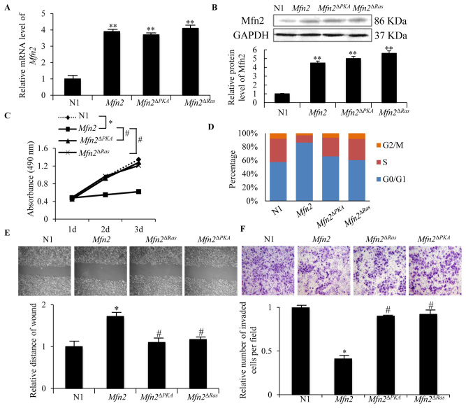
Mitofusin 2 (Mfn2) is expressed in numerous human tissues and serves a pivotal role in cell proliferation. However, Mfn2 is considered as an anti-tumor gene, and is silenced in human malignant tumors, including those of breast cancer. However, the mechanisms contributing to Mfn2 silencing and the mechanism of its anti-tumor function in breast cancer remain unclear. In the present study, hypoexpression of Mfn2, and hypermethylation of its promoter, was confirmed in human breast cancer cells and in breast cancer tissues by reverse transcription-quantitative polymerase chain reaction (PCR) and methylation specific PCR, respectively. Chemical demethylation treatment with 5-aza-2'-deoxycytidine upregulated the mRNA expression level of Mfn2 in MCF-7 cells in a dose-dependent manner. In addition, overexpression of Mfn2 repressed the proliferation, migration and invasion of MCF-7 cells, mediated by inhibition of the Ras-extracellular signal-regulated kinase (ERK)1/2 signaling pathway. However, overexpression of Mfn2 with deletion of the p21Ras motif (Mfn2ΔRas) and protein kinase A (PKA) phosphorylation site (Mfn2ΔPKA) partially reduced the anti-tumor function of Mfn2, and inhibited the Ras-ERK1/2 signaling pathway. Taken together, the present study confirmed the anti-tumor effects of Mfn2 in human breast cancer and clarified that the mechanism of its anti-tumor functions includes promoter DNA methylation, the P21Ras binding site and PKA phosphorylation.
Keywords: DNA methylation; Ras-extracellular signal-regulated kinase 1/2 signaling pathway; human breast cancer; mitofusin 2.
Figures


Similar articles
-
Jixuecao (Herba Centellae Asiaticae) alleviates mesangial cell proliferation in IgA nephropathy by inducing mitofusin 2 expression.J Tradit Chin Med. 2019 Jun;39(3):346-355. J Tradit Chin Med. 2019. PMID: 32186007
-
Function and regulatory mechanisms of the candidate tumor suppressor receptor protein tyrosine phosphatase gamma (PTPRG) in breast cancer cells.Anticancer Res. 2010 Jun;30(6):1937-46. Anticancer Res. 2010. PMID: 20651337
-
Pro-apoptotic and anti-proliferative effects of mitofusin-2 via PI3K/Akt signaling in breast cancer cells.Oncol Lett. 2015 Dec;10(6):3816-3822. doi: 10.3892/ol.2015.3748. Epub 2015 Sep 25. Oncol Lett. 2015. PMID: 26788214 Free PMC article.
-
DNA methylation of claudin-6 promotes breast cancer cell migration and invasion by recruiting MeCP2 and deacetylating H3Ac and H4Ac.J Exp Clin Cancer Res. 2016 Jul 26;35(1):120. doi: 10.1186/s13046-016-0396-x. J Exp Clin Cancer Res. 2016. PMID: 27461117 Free PMC article.
-
Gene methylation in gastric cancer.Clin Chim Acta. 2013 Sep 23;424:53-65. doi: 10.1016/j.cca.2013.05.002. Epub 2013 May 10. Clin Chim Acta. 2013. PMID: 23669186 Review.
Cited by
-
Mfn2 inhibits proliferation and cell-cycle in Hela cells via Ras-NF-κB signal pathway.Cancer Cell Int. 2019 Jul 29;19:197. doi: 10.1186/s12935-019-0916-9. eCollection 2019. Cancer Cell Int. 2019. PMID: 31384172 Free PMC article.
-
The integrated stress response is tumorigenic and constitutes a therapeutic liability in KRAS-driven lung cancer.Nat Commun. 2021 Jul 30;12(1):4651. doi: 10.1038/s41467-021-24661-0. Nat Commun. 2021. PMID: 34330898 Free PMC article.
-
MFN2 suppresses the accumulation of lipid droplets and the progression of clear cell renal cell carcinoma.Cancer Sci. 2024 Jun;115(6):1791-1807. doi: 10.1111/cas.16151. Epub 2024 Mar 13. Cancer Sci. 2024. PMID: 38480904 Free PMC article.
-
Mitochondria, Oxidative Stress, cAMP Signalling and Apoptosis: A Crossroads in Lymphocytes of Multiple Sclerosis, a Possible Role of Nutraceutics.Antioxidants (Basel). 2020 Dec 28;10(1):21. doi: 10.3390/antiox10010021. Antioxidants (Basel). 2020. PMID: 33379309 Free PMC article. Review.
-
The mitochondrial fusion-associated protein MFN2 can be used as a novel prognostic molecule for clear cell renal cell carcinoma.BMC Cancer. 2023 Oct 16;23(1):986. doi: 10.1186/s12885-023-11419-8. BMC Cancer. 2023. PMID: 37845657 Free PMC article.
References
LinkOut - more resources
Full Text Sources
Other Literature Sources
Research Materials
Miscellaneous