In vitro study on role of σB protein in avian reovirus pathogenesis
- PMID: 29731966
- PMCID: PMC5929409
- DOI: 10.18632/oncotarget.24668
In vitro study on role of σB protein in avian reovirus pathogenesis
Abstract
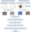
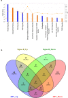
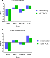
Avian reoviruses, members of Orthoreovirus genus was known to cause diseases like tenosynovitis, runting-stunting syndrome in chickens. Among eight structural proteins, the proteins of S-class are mainly associated with viral arthritis but the significance of σB protein in arthritis is not established till date. In this infection pathological condition together with infection of joints often leads to arthritis because joints consists of cartilage which forms lubricating surface between two bones, and has limited metabolic, replicative and repair capacity. To establish the role of σB protein in arthritis, an in-vitro microarray study was conducted consisting four groups viz. virus infected and control; pDsRed-Express-N1-σB and empty pDs-Red transfected, CEF cells. With cut-off value as FC ≥2, p value <0.05, 6709 and 4026 numbers of DEGs in virus and σB, respectively were identified. The Ingenuity Pathway Analysis gave an idea about the involvement of σB protein in "osteoarthritis pathway", which was activated with z-score with 3.151. The pathway "Role of IL-17A in arthritis pathway" was also enriched with -log (p-value) 1.64. Among total 122 genes involved in osteoarthritis pathway, 28 upregulated and 11 downregulated DEGs were common to both virus and σB treated cells. Moreover, 14 upregulated and 7 downregulated were unique in σB transfected cells. Using qRT-PCR for IL-1B, BMP2, SMAD1, SPP1 genes, the microarray data was validated. We concluded that during ARV infection σB protein, if not fully partially leads to molecular alteration of various genes of host orchestrating the different molecular pattern in joints, leading to tenosynovitis syndrome.
Keywords: S3 gene; arthritis; avian reovirus; microarray; σB protein.
Conflict of interest statement
CONFLICTS OF INTEREST Authors declare that they have no conflicts of interest and provide the informed consent.
Figures


Similar articles
-
Construction of recombinant Marek's disease virus co-expressing σB and σC of avian reoviruses.Front Vet Sci. 2024 Sep 5;11:1461116. doi: 10.3389/fvets.2024.1461116. eCollection 2024. Front Vet Sci. 2024. PMID: 39301286 Free PMC article.
-
Antigenic analysis of monoclonal antibodies against different epitopes of σB protein of avian reovirus.PLoS One. 2013 Nov 27;8(11):e81533. doi: 10.1371/journal.pone.0081533. eCollection 2013. PLoS One. 2013. PMID: 24312314 Free PMC article.
-
Baculovirus surface display of sigmaC and sigmaB proteins of avian reovirus and immunogenicity of the displayed proteins in a mouse model.Vaccine. 2008 Nov 25;26(50):6361-7. doi: 10.1016/j.vaccine.2008.09.008. Epub 2008 Sep 20. Vaccine. 2008. PMID: 18809448
-
Development of ELISA kits for antibodies against avian reovirus using the sigmaC and sigmaB proteins expressed in the methyltropic yeast Pichia pastoris.J Virol Methods. 2010 Feb;163(2):169-74. doi: 10.1016/j.jviromet.2009.07.009. Epub 2009 Jul 28. J Virol Methods. 2010. PMID: 19643143
-
Antigenic analysis monoclonal antibodies against different epitopes of σB protein of Muscovy duck reovirus.Virus Res. 2012 Feb;163(2):546-51. doi: 10.1016/j.virusres.2011.12.006. Epub 2011 Dec 16. Virus Res. 2012. PMID: 22197425
Cited by
-
Wnt14 regulates the cellular inflammation induced by avian reovirus and interacts with the viral σB protein.Poult Sci. 2025 Jun 26;104(9):105493. doi: 10.1016/j.psj.2025.105493. Online ahead of print. Poult Sci. 2025. PMID: 40592292 Free PMC article.
References
-
- Gouvea VS, Schnitzer TJ. Polymorphism of the genomic RNAs among the avian reoviruses. J Gen Virol. 1982;61((Pt l)):87–91. - PubMed
-
- Wickramasinghe R, Meanger J, Enriquez CE, Wilcox GE. Avian reovirus proteins associated with neutralization of virus infectivity. Virology. 1993;194:688–96. - PubMed
-
- Glass SE, Naqi SA, Hall CF, Kerr KM. Isolation and characterization of a virus associated with arthritis of chickens. Avian Dis. 1973;17:415–24. - PubMed
-
- Ni Y, Ramig RF. Characterization of avian reovirus-induced cell fusion: the role of viral structural proteins. Virology. 1993;194:705–14. - PubMed
LinkOut - more resources
Full Text Sources
Other Literature Sources
Molecular Biology Databases
Research Materials
Miscellaneous

